 Movie
Movie Controller

[English] 日本語
 Yorodumi
- PDB-3unc: Crystal Structure of Bovine Milk Xanthine Dehydrogenase to 1.65A ... -
+ Open data

- Basic information
| Entry | Database: PDB / ID: 3unc | ||||||
|---|---|---|---|---|---|---|---|
| Title | Crystal Structure of Bovine Milk Xanthine Dehydrogenase to 1.65A Resolution | ||||||
 Components Components | Xanthine dehydrogenase/oxidase | ||||||
 Keywords Keywords | OXIDOREDUCTASE / Xanthine Dehydrogenase | ||||||
| Function / homology |  Function and homology information Function and homology informationxanthine dehydrogenase complex / xanthine dehydrogenase / xanthine oxidase / xanthine oxidase activity / xanthine catabolic process / xanthine dehydrogenase activity / molybdenum ion binding / molybdopterin cofactor binding / FAD binding / 2 iron, 2 sulfur cluster binding ...xanthine dehydrogenase complex / xanthine dehydrogenase / xanthine oxidase / xanthine oxidase activity / xanthine catabolic process / xanthine dehydrogenase activity / molybdenum ion binding / molybdopterin cofactor binding / FAD binding / 2 iron, 2 sulfur cluster binding / flavin adenine dinucleotide binding / peroxisome / iron ion binding / protein homodimerization activity / : Similarity search - Function | ||||||
| Biological species |  | ||||||
| Method |  X-RAY DIFFRACTION / SYNCHROTRON / MOLECULAR REPLACEMENT / Resolution: 1.65 Å X-RAY DIFFRACTION / SYNCHROTRON / MOLECULAR REPLACEMENT / Resolution: 1.65 Å | ||||||
 Authors Authors | Eger, B.T. / Okamoto, K. / Nishino, T. / Pai, E.F. | ||||||
 Citation Citation | Journal: J.Am.Chem.Soc. / Year: 2012 Title: Protein conformational gating of enzymatic activity in xanthine oxidoreductase. Authors: Ishikita, H. / Eger, B.T. / Okamoto, K. / Nishino, T. / Pai, E.F. | ||||||
| History |
|
- Structure visualization
| Structure viewer | Molecule: MolmilJmol/JSmol |
|---|
- Downloads & links
-Download
| PDBx/mmCIF format | 3unc.cif.gz | 565.7 KB | Display | PDBx/mmCIF format |
|---|---|---|---|---|
| PDB format | pdb3unc.ent.gz | 450.5 KB | Display | PDB format |
| PDBx/mmJSON format | 3unc.json.gz | Tree view | PDBx/mmJSON format | |
| Others |  Other downloads Other downloads |
-Validation report
| Arichive directory | https://data.pdbj.org/pub/pdb/validation_reports/un/3uncftp://data.pdbj.org/pub/pdb/validation_reports/un/3unc | HTTPS FTP |
|---|
-Related structure data
| Related structure data |  3ax7C  3ax9C  3unaC  3uniC  1fo4S S: Starting model for refinement C: citing same article ( |
|---|---|
| Similar structure data |


-Links
 PDBj
PDBj


- Assembly
| Deposited unit | 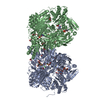
| ||||||||
|---|---|---|---|---|---|---|---|---|---|
| 1 |
| ||||||||
| Unit cell |
|
-Components
-Protein , 1 types, 2 molecules AB
| #1: Protein | Mass: 146970.078 Da / Num. of mol.: 2 / Source method: isolated from a natural source / Details: Isolated from Milk / Source: (natural) References: UniProt: P80457, xanthine dehydrogenase, xanthine oxidase |
|---|
-Non-polymers , 9 types, 2006 molecules 


| #2: Chemical | ChemComp-FES /  Mass: 175.820 Da / Num. of mol.: 4 / Source method: obtained synthetically / Formula: Fe2S2 Mass: 175.820 Da / Num. of mol.: 4 / Source method: obtained synthetically / Formula: Fe2S2#3: Chemical |  Mass: 395.352 Da / Num. of mol.: 2 / Source method: obtained synthetically / Formula: C10H14N5O6PS2 Mass: 395.352 Da / Num. of mol.: 2 / Source method: obtained synthetically / Formula: C10H14N5O6PS2#4: Chemical |  Mass: 161.012 Da / Num. of mol.: 2 / Source method: obtained synthetically / Formula: HMoO2S Mass: 161.012 Da / Num. of mol.: 2 / Source method: obtained synthetically / Formula: HMoO2S#5: Chemical |  Mass: 785.550 Da / Num. of mol.: 2 / Source method: obtained synthetically / Formula: C27H33N9O15P2 / Comment: FAD*YM Mass: 785.550 Da / Num. of mol.: 2 / Source method: obtained synthetically / Formula: C27H33N9O15P2 / Comment: FAD*YM#6: Chemical |  Mass: 138.121 Da / Num. of mol.: 2 / Source method: obtained synthetically / Formula: C7H6O3 Mass: 138.121 Da / Num. of mol.: 2 / Source method: obtained synthetically / Formula: C7H6O3#7: Chemical | ChemComp-GOL /  Mass: 92.094 Da / Num. of mol.: 24 / Source method: obtained synthetically / Formula: C3H8O3 Mass: 92.094 Da / Num. of mol.: 24 / Source method: obtained synthetically / Formula: C3H8O3#8: Chemical |  Mass: 60.009 Da / Num. of mol.: 2 / Source method: obtained synthetically / Formula: CO3 Mass: 60.009 Da / Num. of mol.: 2 / Source method: obtained synthetically / Formula: CO3#9: Chemical |  Mass: 40.078 Da / Num. of mol.: 2 / Source method: obtained synthetically / Formula: Ca Mass: 40.078 Da / Num. of mol.: 2 / Source method: obtained synthetically / Formula: Ca#10: Water | ChemComp-HOH / | Mass: 18.015 Da / Num. of mol.: 1966 / Source method: isolated from a natural source / Formula: H2O |
|---|
-Experimental details
-Experiment
| Experiment | Method: X-RAY DIFFRACTION / Number of used crystals: 1 |
|---|
- Sample preparation
| Crystal | Density Matthews: 2.63 Å3/Da / Density % sol: 53.16 % |
|---|---|
| Crystal grow | Temperature: 293 K / Method: sitting drop batch slide / pH: 7.5 Details: 12 mg/ml Xanthine Dehydrogenase (from Bovine Milk), 8 % Polyethylene Glycol 4000, 30 % Glycerol, 5 mM Dithiothreitol, 0.5 mM Sodium Salicylate, 0.2 m Ethylenediaminetetraacetic Acid, 16.65 ...Details: 12 mg/ml Xanthine Dehydrogenase (from Bovine Milk), 8 % Polyethylene Glycol 4000, 30 % Glycerol, 5 mM Dithiothreitol, 0.5 mM Sodium Salicylate, 0.2 m Ethylenediaminetetraacetic Acid, 16.65 mM Sodium Pyrophosphate (pH 8.5), 25 mM Potasium Phosphate (pH 6.5). , Sitting Drop Batch Slide, temperature 293.0K |
-Data collection
| Diffraction | Mean temperature: 93 K |
|---|---|
| Diffraction source | Source: SYNCHROTRON / Site: SPring-8  / Beamline: BL44B2 / Wavelength: 1 Å / Beamline: BL44B2 / Wavelength: 1 Å |
| Detector | Type: ADSC QUANTUM 210 / Detector: CCD / Date: Mar 14, 2001 |
| Radiation | Protocol: SINGLE WAVELENGTH / Monochromatic (M) / Laue (L): M / Scattering type: x-ray |
| Radiation wavelength | Wavelength: 1 Å / Relative weight: 1 |
| Reflection | Resolution: 1.65→20 Å / Num. all: 364190 / Num. obs: 363495 / % possible obs: 99.9 % / Observed criterion σ(F): -3 / Observed criterion σ(I): -3 / Redundancy: 4.3 % / Rmerge(I) obs: 0.053 / Net I/σ(I): 25.5 |
| Reflection shell | Resolution: 1.65→1.67 Å / Redundancy: 3.8 % / Rmerge(I) obs: 0.454 / Num. unique all: 14437 / % possible all: 99.9 |
- Processing
| Software |
| ||||||||||||||||||||
|---|---|---|---|---|---|---|---|---|---|---|---|---|---|---|---|---|---|---|---|---|---|
| Refinement | Method to determine structure: MOLECULAR REPLACEMENT Starting model: PBD Entry 1FO4 Resolution: 1.65→20 Å / Cross valid method: shells / σ(F): 0 / σ(I): 0 / Stereochemistry target values: Engh & Huber
| ||||||||||||||||||||
| Displacement parameters |
| ||||||||||||||||||||
| Refinement step | Cycle: LAST / Resolution: 1.65→20 Å
| ||||||||||||||||||||
| Refine LS restraints |
|
