 Movie
Movie Controller

[English] 日本語
 Yorodumi
- PDB-1fo4: CRYSTAL STRUCTURE OF XANTHINE DEHYDROGENASE ISOLATED FROM BOVINE MILK -
+ Open data

- Basic information
| Entry | Database: PDB / ID: 1fo4 | ||||||
|---|---|---|---|---|---|---|---|
| Title | CRYSTAL STRUCTURE OF XANTHINE DEHYDROGENASE ISOLATED FROM BOVINE MILK | ||||||
 Components Components | XANTHINE DEHYDROGENASE | ||||||
 Keywords Keywords | OXIDOREDUCTASE / Xanthine Dehydrogenase / FAD / molybdopterin / 2Fe-2S iron sulfur centers / salicylate | ||||||
| Function / homology |  Function and homology information Function and homology informationxanthine dehydrogenase complex / xanthine dehydrogenase / xanthine oxidase / xanthine oxidase activity / xanthine catabolic process / xanthine dehydrogenase activity / molybdenum ion binding / molybdopterin cofactor binding / FAD binding / 2 iron, 2 sulfur cluster binding ...xanthine dehydrogenase complex / xanthine dehydrogenase / xanthine oxidase / xanthine oxidase activity / xanthine catabolic process / xanthine dehydrogenase activity / molybdenum ion binding / molybdopterin cofactor binding / FAD binding / 2 iron, 2 sulfur cluster binding / flavin adenine dinucleotide binding / peroxisome / iron ion binding / protein homodimerization activity / : Similarity search - Function | ||||||
| Biological species |  | ||||||
| Method |  X-RAY DIFFRACTION / SYNCHROTRON / Resolution: 2.1 Å X-RAY DIFFRACTION / SYNCHROTRON / Resolution: 2.1 Å | ||||||
 Authors Authors | Enroth, C. / Eger, B.T. / Okamoto, K. / Nishino, T. / Nishino, T. / Pai, E.F. | ||||||
 Citation Citation | Journal: Proc.Natl.Acad.Sci.USA / Year: 2000 Title: Crystal structures of bovine milk xanthine dehydrogenase and xanthine oxidase: structure-based mechanism of conversion. Authors: Enroth, C. / Eger, B.T. / Okamoto, K. / Nishino, T. / Nishino, T. / Pai, E.F. | ||||||
| History |
|
- Structure visualization
| Structure viewer | Molecule: MolmilJmol/JSmol |
|---|
- Downloads & links
-Download
| PDBx/mmCIF format | 1fo4.cif.gz | 562.4 KB | Display | PDBx/mmCIF format |
|---|---|---|---|---|
| PDB format | pdb1fo4.ent.gz | 447.1 KB | Display | PDB format |
| PDBx/mmJSON format | 1fo4.json.gz | Tree view | PDBx/mmJSON format | |
| Others |  Other downloads Other downloads |
-Validation report
| Arichive directory | https://data.pdbj.org/pub/pdb/validation_reports/fo/1fo4ftp://data.pdbj.org/pub/pdb/validation_reports/fo/1fo4 | HTTPS FTP |
|---|
-Related structure data


-Links
 PDBj
PDBj


- Assembly
| Deposited unit | 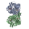
| ||||||||
|---|---|---|---|---|---|---|---|---|---|
| 1 |
| ||||||||
| Unit cell |
| ||||||||
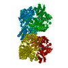
| Details | The biological assmbly is a dimer constructed from a single chain in each biological monomer. The monomer is divided into 3 chains due to 2 missing loops of electron density. The dimer is generated by a two-fold. |
-Components
-Protein , 1 types, 2 molecules AB
| #1: Protein | Mass: 146993.141 Da / Num. of mol.: 2 / Source method: isolated from a natural source / Source: (natural) |
|---|
-Non-polymers , 8 types, 2065 molecules 


| #2: Chemical |  Mass: 40.078 Da / Num. of mol.: 2 / Source method: obtained synthetically / Formula: Ca Mass: 40.078 Da / Num. of mol.: 2 / Source method: obtained synthetically / Formula: Ca#3: Chemical | ChemComp-FES /  Mass: 175.820 Da / Num. of mol.: 4 / Source method: obtained synthetically / Formula: Fe2S2 Mass: 175.820 Da / Num. of mol.: 4 / Source method: obtained synthetically / Formula: Fe2S2#4: Chemical |  Mass: 395.352 Da / Num. of mol.: 2 / Source method: obtained synthetically / Formula: C10H14N5O6PS2 Mass: 395.352 Da / Num. of mol.: 2 / Source method: obtained synthetically / Formula: C10H14N5O6PS2#5: Chemical |  Mass: 161.012 Da / Num. of mol.: 2 / Source method: obtained synthetically / Formula: HMoO2S Mass: 161.012 Da / Num. of mol.: 2 / Source method: obtained synthetically / Formula: HMoO2S#6: Chemical |  Mass: 138.121 Da / Num. of mol.: 2 / Source method: obtained synthetically / Formula: C7H6O3 Mass: 138.121 Da / Num. of mol.: 2 / Source method: obtained synthetically / Formula: C7H6O3#7: Chemical |  Mass: 785.550 Da / Num. of mol.: 2 / Source method: obtained synthetically / Formula: C27H33N9O15P2 / Comment: FAD*YM Mass: 785.550 Da / Num. of mol.: 2 / Source method: obtained synthetically / Formula: C27H33N9O15P2 / Comment: FAD*YM#8: Chemical | ChemComp-GOL /  Mass: 92.094 Da / Num. of mol.: 4 / Source method: obtained synthetically / Formula: C3H8O3 Mass: 92.094 Da / Num. of mol.: 4 / Source method: obtained synthetically / Formula: C3H8O3#9: Water | ChemComp-HOH / | Mass: 18.015 Da / Num. of mol.: 2047 / Source method: isolated from a natural source / Formula: H2O |
|---|
-Experimental details
-Experiment
| Experiment | Method: X-RAY DIFFRACTION / Number of used crystals: 1 |
|---|
- Sample preparation
| Crystal | Density Matthews: 2.66 Å3/Da / Density % sol: 53.75 % |
|---|---|
| Crystal grow | Temperature: 295 K / Method: vapor diffusion, sitting drop / pH: 7.5 Details: PEG 4000, DTT, glycerol, KPi, NaPPi, salicylate, EDTA, pH 7.5, VAPOR DIFFUSION, SITTING DROP, temperature 295.0K |
| Crystal grow | *PLUS Method: unknown / Details: unpublished data |
-Data collection
| Diffraction | Mean temperature: 100 K |
|---|---|
| Diffraction source | Source: SYNCHROTRON / Site: NSLS  / Beamline: X8C / Wavelength: 0.97895 / Beamline: X8C / Wavelength: 0.97895 |
| Detector | Type: ADSC QUANTUM 4 / Detector: CCD / Date: Feb 7, 1999 |
| Radiation | Protocol: SINGLE WAVELENGTH / Monochromatic (M) / Laue (L): M / Scattering type: x-ray |
| Radiation wavelength | Wavelength: 0.97895 Å / Relative weight: 1 |
| Reflection | Resolution: 2.1→24.9 Å / Num. all: 176889 / Num. obs: 154198 / % possible obs: 87.2 % / Observed criterion σ(F): 0 / Observed criterion σ(I): -3 / Redundancy: 2.8 % / Biso Wilson estimate: 14.2 Å2 / Rmerge(I) obs: 0.095 / Net I/σ(I): 11.9 |
| Reflection shell | Resolution: 2.1→2.14 Å / Redundancy: 2.1 % / Rmerge(I) obs: 0.387 / Num. unique all: 5988 / % possible all: 67.8 |
| Reflection | *PLUS Num. obs: 7710 / Num. measured all: 154198 |
| Reflection shell | *PLUS % possible obs: 67.8 % |
- Processing
| Software |
| |||||||||||||||||||||||||
|---|---|---|---|---|---|---|---|---|---|---|---|---|---|---|---|---|---|---|---|---|---|---|---|---|---|---|
| Refinement | Resolution: 2.1→25 Å / σ(F): 0 / σ(I): 0 / Stereochemistry target values: Engh & Huber
| |||||||||||||||||||||||||
| Refinement step | Cycle: LAST / Resolution: 2.1→25 Å
| |||||||||||||||||||||||||
| Refine LS restraints |
| |||||||||||||||||||||||||
| Software | *PLUS Name: CNS / Classification: refinement | |||||||||||||||||||||||||
| Refinement | *PLUS | |||||||||||||||||||||||||
| Solvent computation | *PLUS | |||||||||||||||||||||||||
| Displacement parameters | *PLUS Biso mean: 18.8 Å2 |
