 Movie
Movie Controller

[English] 日本語
 Yorodumi
- PDB-3bdj: Crystal Structure of Bovine Milk Xanthine Dehydrogenase with a Co... -
+ Open data

- Basic information
| Entry | Database: PDB / ID: 3bdj | ||||||
|---|---|---|---|---|---|---|---|
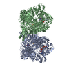
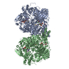
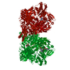
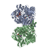
| Title | Crystal Structure of Bovine Milk Xanthine Dehydrogenase with a Covalently Bound Oxipurinol Inhibitor | ||||||
 Components Components | Xanthine dehydrogenase/oxidase | ||||||
 Keywords Keywords | OXIDOREDUCTASE / OXYPURINOL / OXIPURINOL / ALLOXANTHINE / ALLOPURINOL / COVALENTLY BOUND INHIBITOR / XANTHINE OXIDASE / XANTHINE OXIDOREDUCTASE / XANTHINE DEHYDROGENASE / FAD / FLAVOPROTEIN / IRON-SULFUR / MOLYBDOPTERIN / PEROXISOME | ||||||
| Function / homology |  Function and homology information Function and homology informationxanthine dehydrogenase complex / xanthine dehydrogenase / xanthine oxidase / xanthine oxidase activity / xanthine catabolic process / xanthine dehydrogenase activity / molybdenum ion binding / molybdopterin cofactor binding / FAD binding / 2 iron, 2 sulfur cluster binding ...xanthine dehydrogenase complex / xanthine dehydrogenase / xanthine oxidase / xanthine oxidase activity / xanthine catabolic process / xanthine dehydrogenase activity / molybdenum ion binding / molybdopterin cofactor binding / FAD binding / 2 iron, 2 sulfur cluster binding / flavin adenine dinucleotide binding / peroxisome / iron ion binding / protein homodimerization activity / : Similarity search - Function | ||||||
| Biological species |  | ||||||
| Method |  X-RAY DIFFRACTION / SYNCHROTRON / MOLECULAR REPLACEMENT / Resolution: 2 Å X-RAY DIFFRACTION / SYNCHROTRON / MOLECULAR REPLACEMENT / Resolution: 2 Å | ||||||
 Authors Authors | Eger, B.T. / Okamoto, K. / Nishino, T. / Pai, E.F. / Nishino, T. | ||||||
 Citation Citation | Journal: Nucleosides Nucleotides Nucleic Acids / Year: 2008 Title: Mechanism of inhibition of xanthine oxidoreductase by allopurinol: crystal structure of reduced bovine milk xanthine oxidoreductase bound with oxipurinol. Authors: Okamoto, K. / Eger, B.T. / Nishino, T. / Pai, E.F. / Nishino, T. #1: Journal: Structure / Year: 2002Title: Crystal Structures of the Active and Alloxanthine-inhibited forms of Xanthine Dehydrogenase from Rhodobacter Capsulatusa Authors: Truglio, J.J. / Theis, K. / Leimkuhler, S. / Rappa, R. / Rajagopalan, K.V. / Kisker, C. #2: Journal: Proc.Natl.Acad.Sci.USA / Year: 2004Title: The Crystal Structure of Xanthine Oxidoreductase during catalysis: Implications for reaction mechanism and enzyme inhibition Authors: Okamoto, K. / Matsumoto, K. / Hille, R. / Eger, B.T. / Pai, E.F. / Nishino, T. #3: Journal: J.PHARMACOL.EXP.THER. / Year: 2004Title: Y-700[1-[3-CYANO-4-(2,2-DIMETHYLPROPOXY)PHENYL]-1H-PYRAZOLE-4-CARBOXYLIC ACID]: A Potent Xanthine Oxidoreductase Inhibitor with Hepatic excretion Authors: Fukunari, A. / Okamoto, K. / Nishino, T. / Eger, B.T. / Pai, E.F. / Kamezawa, M. / Yamada, I. / Kato, N. #4: Journal: J.Biol.Chem. / Year: 2003Title: An extremely potent inhibitor of Xanthine Oxidoreductase Crystal Structure of the enzyme-inhibitor complex and Mechanism of Inhibition Authors: Okamoto, K. / Eger, B.T. / Nishino, T. / Kondo, S. / Pai, E.F. / Nishino, T. | ||||||
| History |
|
- Structure visualization
| Structure viewer | Molecule: MolmilJmol/JSmol |
|---|
- Downloads & links
-Download
| PDBx/mmCIF format | 3bdj.cif.gz | 545.1 KB | Display | PDBx/mmCIF format |
|---|---|---|---|---|
| PDB format | pdb3bdj.ent.gz | 434.6 KB | Display | PDB format |
| PDBx/mmJSON format | 3bdj.json.gz | Tree view | PDBx/mmJSON format | |
| Others |  Other downloads Other downloads |
-Validation report
| Arichive directory | https://data.pdbj.org/pub/pdb/validation_reports/bd/3bdjftp://data.pdbj.org/pub/pdb/validation_reports/bd/3bdj | HTTPS FTP |
|---|
-Related structure data
| Related structure data |  1fo4S S: Starting model for refinement |
|---|---|
| Similar structure data |


-Links
 PDBj
PDBj


- Assembly
| Deposited unit | 
| ||||||||
|---|---|---|---|---|---|---|---|---|---|
| 1 |
| ||||||||
| Unit cell |
|
-Components
-Protein , 1 types, 2 molecules AB
| #1: Protein | Mass: 146993.141 Da / Num. of mol.: 2 / Source method: isolated from a natural source / Source: (natural) References: UniProt: P80457, xanthine oxidase, xanthine dehydrogenase |
|---|
-Non-polymers , 9 types, 1417 molecules 


| #2: Chemical |  Mass: 40.078 Da / Num. of mol.: 2 / Source method: obtained synthetically / Formula: Ca Mass: 40.078 Da / Num. of mol.: 2 / Source method: obtained synthetically / Formula: Ca#3: Chemical | ChemComp-FES /  Mass: 175.820 Da / Num. of mol.: 4 / Source method: obtained synthetically / Formula: Fe2S2 Mass: 175.820 Da / Num. of mol.: 4 / Source method: obtained synthetically / Formula: Fe2S2#4: Chemical |  Mass: 395.352 Da / Num. of mol.: 2 / Source method: obtained synthetically / Formula: C10H14N5O6PS2 Mass: 395.352 Da / Num. of mol.: 2 / Source method: obtained synthetically / Formula: C10H14N5O6PS2#5: Chemical |  Mass: 145.012 Da / Num. of mol.: 2 / Source method: obtained synthetically / Formula: HMoOS Mass: 145.012 Da / Num. of mol.: 2 / Source method: obtained synthetically / Formula: HMoOS#6: Chemical |  Mass: 785.550 Da / Num. of mol.: 2 / Source method: obtained synthetically / Formula: C27H33N9O15P2 / Comment: FAD*YM Mass: 785.550 Da / Num. of mol.: 2 / Source method: obtained synthetically / Formula: C27H33N9O15P2 / Comment: FAD*YM#7: Chemical |  Mass: 152.111 Da / Num. of mol.: 2 / Source method: obtained synthetically / Formula: C5H4N4O2 Mass: 152.111 Da / Num. of mol.: 2 / Source method: obtained synthetically / Formula: C5H4N4O2#8: Chemical | ChemComp-GOL /  Mass: 92.094 Da / Num. of mol.: 7 / Source method: obtained synthetically / Formula: C3H8O3 Mass: 92.094 Da / Num. of mol.: 7 / Source method: obtained synthetically / Formula: C3H8O3#9: Chemical |  Mass: 60.009 Da / Num. of mol.: 2 / Source method: obtained synthetically / Formula: CO3 Mass: 60.009 Da / Num. of mol.: 2 / Source method: obtained synthetically / Formula: CO3#10: Water | ChemComp-HOH / | Mass: 18.015 Da / Num. of mol.: 1394 / Source method: isolated from a natural source / Formula: H2O |
|---|
-Details
| Sequence details | UNP ENTRY P80457 HAS A CONFLICT ASP TO HIS AT RESIDUE 552 |
|---|
-Experimental details
-Experiment
| Experiment | Method: X-RAY DIFFRACTION / Number of used crystals: 1 |
|---|
- Sample preparation
| Crystal | Density Matthews: 2.61 Å3/Da / Density % sol: 52.94 % |
|---|---|
| Crystal grow | Temperature: 295 K / Method: vapor diffusion, sitting drop / pH: 7.5 Details: PEG 4000, DTT, GLYCEROL, KPI, NAPPI, EDTA, pH 7.50, VAPOR DIFFUSION, SITTING DROP, temperature 295K |
-Data collection
| Diffraction | Mean temperature: 100 K |
|---|---|
| Diffraction source | Source: SYNCHROTRON / Site: SPring-8  / Beamline: BL40B2 / Wavelength: 1 Å / Beamline: BL40B2 / Wavelength: 1 Å |
| Detector | Type: ADSC QUANTUM 4 / Detector: CCD / Date: Nov 13, 2001 |
| Radiation | Protocol: SINGLE WAVELENGTH / Monochromatic (M) / Laue (L): M / Scattering type: x-ray |
| Radiation wavelength | Wavelength: 1 Å / Relative weight: 1 |
| Reflection | Resolution: 2→20 Å / Num. all: 204186 / Num. obs: 192610 / % possible obs: 94.4 % / Observed criterion σ(F): -3 / Observed criterion σ(I): -3 / Redundancy: 3.2 % / Biso Wilson estimate: 11.8 Å2 / Rmerge(I) obs: 0.072 / Net I/σ(I): 14.5 |
| Reflection shell | Resolution: 2→2.03 Å / Redundancy: 2.4 % / Rmerge(I) obs: 0.409 / Mean I/σ(I) obs: 2.2 / % possible all: 80.9 |
- Processing
| Software |
| ||||||||||||||||||||||||||||||||||||||||||||||||||||||||||||
|---|---|---|---|---|---|---|---|---|---|---|---|---|---|---|---|---|---|---|---|---|---|---|---|---|---|---|---|---|---|---|---|---|---|---|---|---|---|---|---|---|---|---|---|---|---|---|---|---|---|---|---|---|---|---|---|---|---|---|---|---|---|
| Refinement | Method to determine structure: MOLECULAR REPLACEMENT Starting model: PDB entry 1fo4 Resolution: 2→19.96 Å / Isotropic thermal model: RESTRAINED / Cross valid method: THROUGHOUT / σ(F): 0 / Stereochemistry target values: ENGH & HUBER
| ||||||||||||||||||||||||||||||||||||||||||||||||||||||||||||
| Displacement parameters | Biso mean: 21.2 Å2
| ||||||||||||||||||||||||||||||||||||||||||||||||||||||||||||
| Refine analyze |
| ||||||||||||||||||||||||||||||||||||||||||||||||||||||||||||
| Refinement step | Cycle: LAST / Resolution: 2→19.96 Å
| ||||||||||||||||||||||||||||||||||||||||||||||||||||||||||||
| Refine LS restraints |
| ||||||||||||||||||||||||||||||||||||||||||||||||||||||||||||
| LS refinement shell | Resolution: 2→2.13 Å / Rfactor Rfree error: 0.017
|
