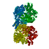
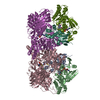
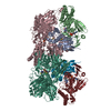
[English] 日本語
Yorodumi
- PDB-1v97: Crystal Structure of Bovine Milk Xanthine Dehydrogenase FYX-051 b... -

+
Open data


ID or keywords:

Loading...

-
Basic information

Entry
Database: PDB / ID: 1v97
TitleCrystal Structure of Bovine Milk Xanthine Dehydrogenase FYX-051 bound form
ComponentsXanthine dehydrogenase
KeywordsOXIDOREDUCTASE / xanthine dehydrogenase / molybdopterin / FYX-051 / reaction intermediate
Function / homology
Function and homology information


xanthine dehydrogenase complex / xanthine dehydrogenase / xanthine oxidase / xanthine oxidase activity / xanthine catabolic process / xanthine dehydrogenase activity / molybdenum ion binding / molybdopterin cofactor binding / FAD binding / 2 iron, 2 sulfur cluster binding ...xanthine dehydrogenase complex / xanthine dehydrogenase / xanthine oxidase / xanthine oxidase activity / xanthine catabolic process / xanthine dehydrogenase activity / molybdenum ion binding / molybdopterin cofactor binding / FAD binding / 2 iron, 2 sulfur cluster binding / peroxisome / flavin adenine dinucleotide binding / iron ion binding / protein homodimerization activity / extracellular space
Similarity search - Function
Xanthine dehydrogenase, small subunit / Oxidoreductase, molybdopterin binding site / Eukaryotic molybdopterin oxidoreductases signature. / Aldehyde oxidase/xanthine dehydrogenase, a/b hammerhead / CO dehydrogenase flavoprotein, C-terminal domain / CO dehydrogenase flavoprotein C-terminal domain / Aldehyde Oxidoreductase; domain 4 / Aldehyde oxidase/xanthine dehydrogenase, molybdopterin binding domain / [2Fe-2S]-binding domain / Molybdopterin dehydrogenase, FAD-binding ...Xanthine dehydrogenase, small subunit / Oxidoreductase, molybdopterin binding site / Eukaryotic molybdopterin oxidoreductases signature. / Aldehyde oxidase/xanthine dehydrogenase, a/b hammerhead / CO dehydrogenase flavoprotein, C-terminal domain / CO dehydrogenase flavoprotein C-terminal domain / Aldehyde Oxidoreductase; domain 4 / Aldehyde oxidase/xanthine dehydrogenase, molybdopterin binding domain / [2Fe-2S]-binding domain / Molybdopterin dehydrogenase, FAD-binding / CO dehydrogenase flavoprotein, C-terminal / CO dehydrogenase flavoprotein, C-terminal domain superfamily / FAD binding domain in molybdopterin dehydrogenase / CO dehydrogenase flavoprotein C-terminal domain / Aldehyde oxidase/xanthine dehydrogenase / Aldehyde oxidase/xanthine dehydrogenase, second molybdopterin binding domain / Molybdopterin cofactor-binding domain / Aldehyde oxidase and xanthine dehydrogenase, a/b hammerhead domain / Aldehyde oxidase/xanthine dehydrogenase, a/b hammerhead / [2Fe-2S]-binding / Aldehyde oxidase/xanthine dehydrogenase, first molybdopterin binding domain / Aldehyde oxidase/xanthine dehydrogenase, a/b hammerhead superfamily / [2Fe-2S]-binding domain superfamily / Aldehyde oxidase/xanthine dehydrogenase, molybdopterin binding domain superfamily / [2Fe-2S] binding domain / Molybdopterin cofactor-binding domain / Aldehyde oxidase and xanthine dehydrogenase, a/b hammerhead domain / Uridine Diphospho-n-acetylenolpyruvylglucosamine Reductase, domain 2 / Uridine Diphospho-n-acetylenolpyruvylglucosamine Reductase; domain 2 / Uridine Diphospho-n-acetylenolpyruvylglucosamine Reductase; domain 3 - #10 / Uridine Diphospho-n-acetylenolpyruvylglucosamine Reductase; domain 3 / FAD-binding, type PCMH, subdomain 1 / Aldehyde Oxidoreductase; domain 3 / 2Fe-2S ferredoxin, iron-sulphur binding site / 2Fe-2S ferredoxin-type iron-sulfur binding region signature. / FAD-binding domain, PCMH-type / PCMH-type FAD-binding domain profile. / 2Fe-2S iron-sulfur cluster binding domain / Beta-grasp domain / FAD-binding, type PCMH, subdomain 2 / FAD-binding, type PCMH-like superfamily / Beta-grasp domain superfamily / Enolase-like; domain 1 / 2Fe-2S ferredoxin-type iron-sulfur binding domain profile. / 2Fe-2S ferredoxin-type iron-sulfur binding domain / 2Fe-2S ferredoxin-like superfamily / Ubiquitin-like (UB roll) / DNA polymerase; domain 1 / Roll / Alpha-Beta Complex / 2-Layer Sandwich / Orthogonal Bundle / Mainly Alpha / Alpha Beta
Similarity search - Domain/homology
ACETIC ACID / FLAVIN-ADENINE DINUCLEOTIDE / FE2/S2 (INORGANIC) CLUSTER / Chem-FYX / DIOXOTHIOMOLYBDENUM(VI) ION / Chem-MTE / Xanthine dehydrogenase/oxidase
Similarity search - Component
Biological speciesBos taurus (cattle)
MethodX-RAY DIFFRACTION / SYNCHROTRON / MOLECULAR REPLACEMENT / Resolution: 1.94 Å
AuthorsOkamoto, K. / Matsumoto, K. / Hille, R. / Eger, B.T. / Pai, E.F. / Nishino, T.
CitationJournal: Proc.Natl.Acad.Sci.USA / Year: 2004
Title: The crystal structure of xanthine oxidoreductase during catalysis: Implications for reaction mechanism and enzyme inhibition.
Authors: Okamoto, K. / Matsumoto, K. / Hille, R. / Eger, B.T. / Pai, E.F. / Nishino, T.
History
DepositionJan 21, 2004Deposition site: PDBJ / Processing site: PDBJ
Revision 1.0Jun 22, 2004Provider: repository / Type: Initial release
Revision 1.1Apr 27, 2008Group: Version format compliance
Revision 1.2Jul 13, 2011Group: Non-polymer description / Version format compliance
Revision 1.3Dec 27, 2023Group: Data collection / Database references / Derived calculations
Category: chem_comp_atom / chem_comp_bond ...chem_comp_atom / chem_comp_bond / database_2 / pdbx_struct_conn_angle / struct_conn / struct_conn_type / struct_site
Item: _database_2.pdbx_DOI / _database_2.pdbx_database_accession ..._database_2.pdbx_DOI / _database_2.pdbx_database_accession / _pdbx_struct_conn_angle.ptnr1_auth_asym_id / _pdbx_struct_conn_angle.ptnr1_auth_comp_id / _pdbx_struct_conn_angle.ptnr1_auth_seq_id / _pdbx_struct_conn_angle.ptnr1_label_asym_id / _pdbx_struct_conn_angle.ptnr1_label_atom_id / _pdbx_struct_conn_angle.ptnr1_label_comp_id / _pdbx_struct_conn_angle.ptnr1_label_seq_id / _pdbx_struct_conn_angle.ptnr2_auth_asym_id / _pdbx_struct_conn_angle.ptnr2_auth_comp_id / _pdbx_struct_conn_angle.ptnr2_auth_seq_id / _pdbx_struct_conn_angle.ptnr2_label_asym_id / _pdbx_struct_conn_angle.ptnr2_label_atom_id / _pdbx_struct_conn_angle.ptnr2_label_comp_id / _pdbx_struct_conn_angle.ptnr3_auth_asym_id / _pdbx_struct_conn_angle.ptnr3_auth_comp_id / _pdbx_struct_conn_angle.ptnr3_auth_seq_id / _pdbx_struct_conn_angle.ptnr3_label_asym_id / _pdbx_struct_conn_angle.ptnr3_label_atom_id / _pdbx_struct_conn_angle.ptnr3_label_comp_id / _pdbx_struct_conn_angle.ptnr3_label_seq_id / _pdbx_struct_conn_angle.value / _struct_conn.conn_type_id / _struct_conn.id / _struct_conn.pdbx_dist_value / _struct_conn.pdbx_leaving_atom_flag / _struct_conn.ptnr1_auth_asym_id / _struct_conn.ptnr1_auth_comp_id / _struct_conn.ptnr1_auth_seq_id / _struct_conn.ptnr1_label_asym_id / _struct_conn.ptnr1_label_atom_id / _struct_conn.ptnr1_label_comp_id / _struct_conn.ptnr1_label_seq_id / _struct_conn.ptnr2_auth_asym_id / _struct_conn.ptnr2_auth_comp_id / _struct_conn.ptnr2_auth_seq_id / _struct_conn.ptnr2_label_asym_id / _struct_conn.ptnr2_label_atom_id / _struct_conn.ptnr2_label_comp_id / _struct_conn.ptnr2_label_seq_id / _struct_conn_type.id / _struct_site.pdbx_auth_asym_id / _struct_site.pdbx_auth_comp_id / _struct_site.pdbx_auth_seq_id

-
Structure visualization

Structure viewerMolecule:
MolmilJmol/JSmol

Downloads & links

-
Assembly

Deposited unit
A: Xanthine dehydrogenase
B: Xanthine dehydrogenase
hetero molecules


Theoretical massNumber of molelcules
Total (without water)298,80726
Polymers293,9862
Non-polymers4,82124
Water37,5072082
1


  • Idetical with deposited unit
  • defined by author&software
TypeNameSymmetry operationNumber
identity operation1_555x,y,z1
Buried area14990 Å2
ΔGint-148 kcal/mol
Surface area86980 Å2
MethodPISA
Unit cell
Length a, b, c (Å)167.987, 124.612, 146.926
Angle α, β, γ (deg.)90.00, 90.99, 90.00
Int Tables number5
Space group name H-MC121

-
Components

-
Protein , 1 types, 2 molecules AB

#1: Protein Xanthine dehydrogenase / / XD


Mass: 146993.141 Da / Num. of mol.: 2 / Source method: isolated from a natural source / Source: (natural) Bos taurus (cattle) / References: UniProt: P80457, 1.1.1.204

-
Non-polymers , 9 types, 2106 molecules

#2: Chemical ChemComp-CA / CALCIUM ION


Mass: 40.078 Da / Num. of mol.: 2 / Source method: obtained synthetically / Formula: Ca
#3: Chemical
ChemComp-FES / FE2/S2 (INORGANIC) CLUSTER / Iron–sulfur cluster


Mass: 175.820 Da / Num. of mol.: 4 / Source method: obtained synthetically / Formula: Fe2S2
#4: Chemical ChemComp-MTE / PHOSPHONIC ACIDMONO-(2-AMINO-5,6-DIMERCAPTO-4-OXO-3,7,8A,9,10,10A-HEXAHYDRO-4H-8-OXA-1,3,9,10-TETRAAZA-ANTHRACEN-7-YLMETHYL)ESTER


Mass: 395.352 Da / Num. of mol.: 2 / Source method: obtained synthetically / Formula: C10H14N5O6PS2
#5: Chemical ChemComp-MOS / DIOXOTHIOMOLYBDENUM(VI) ION


Mass: 161.012 Da / Num. of mol.: 2 / Source method: obtained synthetically / Formula: HMoO2S
#6: Chemical ChemComp-FAD / FLAVIN-ADENINE DINUCLEOTIDE / Flavin adenine dinucleotide


Mass: 785.550 Da / Num. of mol.: 2 / Source method: obtained synthetically / Formula: C27H33N9O15P2 / Comment: FAD*YM
#7: Chemical ChemComp-FYX / 4-(5-PYRIDIN-4-YL-1H-1,2,4-TRIAZOL-3-YL)PYRIDINE-2-CARBONITRILE / 4-[5-PYRIDIN-4-YL-1H-[1,2,4]TRIAZOL-3-YL]-PYRIDINE-2-CARBONITRILE / Topiroxostat


Mass: 248.243 Da / Num. of mol.: 2 / Source method: obtained synthetically / Formula: C13H8N6 / Comment: inhibitor*YM
#8: Chemical ChemComp-ACY / ACETIC ACID / Acetic acid


Mass: 60.052 Da / Num. of mol.: 2 / Source method: obtained synthetically / Formula: C2H4O2
#9: Chemical
ChemComp-GOL / GLYCEROL / GLYCERIN / PROPANE-1,2,3-TRIOL / Glycerol


Mass: 92.094 Da / Num. of mol.: 8 / Source method: obtained synthetically / Formula: C3H8O3
#10: Water ChemComp-HOH / water / Water


Mass: 18.015 Da / Num. of mol.: 2082 / Source method: isolated from a natural source / Formula: H2O

-
Experimental details

-
Experiment

ExperimentMethod: X-RAY DIFFRACTION / Number of used crystals: 1

-
Sample preparation

CrystalDensity Matthews: 2.44 Å3/Da / Density % sol: 49.25 %
Crystal growpH: 7.5 / Details: pH 7.5

-
Data collection

DiffractionMean temperature: 100 K
Diffraction sourceSource: SYNCHROTRON / Site: SPring-8 / Beamline: BL38B1 / Wavelength: 1 Å
DetectorType: ADSC QUANTUM 4 / Detector: CCD / Date: May 28, 2003
RadiationProtocol: SINGLE WAVELENGTH / Monochromatic (M) / Laue (L): M / Scattering type: x-ray
Radiation wavelengthWavelength: 1 Å / Relative weight: 1
ReflectionResolution: 1.94→40 Å / Num. obs: 214439 / % possible obs: 99.9 % / Observed criterion σ(I): -3 / Redundancy: 4.1 % / Rmerge(I) obs: 0.079 / Rsym value: 0.079 / Net I/σ(I): 15.153
Reflection shellResolution: 1.94→1.97 Å / Rmerge(I) obs: 0.396

-
Processing

Software
NameClassification
HKL-2000data collection
SCALEPACKdata scaling
EPMRphasing
CNSrefinement
HKL-2000data reduction
RefinementMethod to determine structure: MOLECULAR REPLACEMENT / Resolution: 1.94→40 Å / σ(F): 0
RfactorNum. reflection% reflectionSelection details
Rfree0.208 6405 3 %RANDOM
Rwork0.178 ---
all0.179 ---
obs0.179 214439 99.9 %-
Refinement stepCycle: LAST / Resolution: 1.94→40 Å
ProteinNucleic acidLigandSolventTotal
Num. atoms20125 0 274 2082 22481
Refine LS restraints
Refine-IDTypeDev ideal
X-RAY DIFFRACTIONc_bond_d0.006
X-RAY DIFFRACTIONc_angle_deg1.4
X-RAY DIFFRACTIONc_dihedral_angle_d
X-RAY DIFFRACTIONc_improper_angle_d

+
About Yorodumi

-
News

-
Feb 9, 2022. New format data for meta-information of EMDB entries

New format data for meta-information of EMDB entries

  • Version 3 of the EMDB header file is now the official format.
  • The previous official version 1.9 will be removed from the archive.

Related info.:EMDB header

External links:wwPDB to switch to version 3 of the EMDB data model

-
Aug 12, 2020. Covid-19 info

Covid-19 info

URL: https://pdbj.org/emnavi/covid19.php

New page: Covid-19 featured information page in EM Navigator.

Related info.:Covid-19 info / Mar 5, 2020. Novel coronavirus structure data

+
Mar 5, 2020. Novel coronavirus structure data

Novel coronavirus structure data

Related info.:Yorodumi Speices / Aug 12, 2020. Covid-19 info

External links:COVID-19 featured content - PDBj / Molecule of the Month (242):Coronavirus Proteases

+
Jan 31, 2019. EMDB accession codes are about to change! (news from PDBe EMDB page)

EMDB accession codes are about to change! (news from PDBe EMDB page)

  • The allocation of 4 digits for EMDB accession codes will soon come to an end. Whilst these codes will remain in use, new EMDB accession codes will include an additional digit and will expand incrementally as the available range of codes is exhausted. The current 4-digit format prefixed with “EMD-” (i.e. EMD-XXXX) will advance to a 5-digit format (i.e. EMD-XXXXX), and so on. It is currently estimated that the 4-digit codes will be depleted around Spring 2019, at which point the 5-digit format will come into force.
  • The EM Navigator/Yorodumi systems omit the EMD- prefix.

Related info.:Q: What is EMD? / ID/Accession-code notation in Yorodumi/EM Navigator

External links:EMDB Accession Codes are Changing Soon! / Contact to PDBj

+
Jul 12, 2017. Major update of PDB

Major update of PDB

  • wwPDB released updated PDB data conforming to the new PDBx/mmCIF dictionary.
  • This is a major update changing the version number from 4 to 5, and with Remediation, in which all the entries are updated.
  • In this update, many items about electron microscopy experimental information are reorganized (e.g. em_software).
  • Now, EM Navigator and Yorodumi are based on the updated data.

External links:wwPDB Remediation / Enriched Model Files Conforming to OneDep Data Standards Now Available in the PDB FTP Archive

-
Yorodumi

Thousand views of thousand structures

  • Yorodumi is a browser for structure data from EMDB, PDB, SASBDB, etc.
  • This page is also the successor to EM Navigator detail page, and also detail information page/front-end page for Omokage search.
  • The word "yorodu" (or yorozu) is an old Japanese word meaning "ten thousand". "mi" (miru) is to see.

Related info.:EMDB / PDB / SASBDB / Comparison of 3 databanks / Yorodumi Search / Aug 31, 2016. New EM Navigator & Yorodumi / Yorodumi Papers / Jmol/JSmol / Function and homology information / Changes in new EM Navigator and Yorodumi

Read more