Mass: 18.015 Da / Num. of mol.: 2029 / Source method: isolated from a natural source / Formula: H2O
-
Details
Nonpolymer details
THE LIGAND URATE LABELED AS URC IS METAL-COORDINATED TO MO COMPOUND MOS, MOLYBDPOTERIN. IT WAS ...THE LIGAND URATE LABELED AS URC IS METAL-COORDINATED TO MO COMPOUND MOS, MOLYBDPOTERIN. IT WAS CHEMICALLY SYNTHESIZED.
Sequence details
THERE IS A SEQUENCE CONFLICT AT THIS POSITION. THE USING DATABASE REFERENCE SEQUENCE REFERS REF 1 OF P80457.
-
Experimental details
-
Experiment
Experiment
Method: X-RAY DIFFRACTION / Number of used crystals: 1
-
Sample preparation
Crystal
Density Matthews: 2.64 Å3/Da / Density % sol: 53.34 %
Resolution: 2.1→34.83 Å / Cor.coef. Fo:Fc: 0.967 / Cor.coef. Fo:Fc free: 0.94 / SU B: 3.8 / SU ML: 0.101 / Cross valid method: THROUGHOUT / ESU R: 0.172 / ESU R Free: 0.157 / Stereochemistry target values: MAXIMUM LIKELIHOOD / Details: HYDROGENS HAVE BEEN ADDED IN THE RIDING POSITIONS
Rfactor
Num. reflection
% reflection
Selection details
Rfree
0.2018
8881
5 %
RANDOM
Rwork
0.15093
-
-
-
obs
0.15347
168559
99.35 %
-
Solvent computation
Ion probe radii: 0.8 Å / Shrinkage radii: 0.8 Å / VDW probe radii: 1.4 Å / Solvent model: MASK
Displacement parameters
Biso mean: 25.134 Å2
Baniso -1
Baniso -2
Baniso -3
1-
0 Å2
0 Å2
0 Å2
2-
-
0 Å2
0 Å2
3-
-
-
0 Å2
Refinement step
Cycle: LAST / Resolution: 2.1→34.83 Å
Protein
Nucleic acid
Ligand
Solvent
Total
Num. atoms
20037
0
316
2029
22382
Refine LS restraints
Refine-ID
Type
Dev ideal
Dev ideal target
Number
X-RAY DIFFRACTION
r_bond_refined_d
0.023
0.022
20812
X-RAY DIFFRACTION
r_bond_other_d
X-RAY DIFFRACTION
r_angle_refined_deg
2.025
1.984
28220
X-RAY DIFFRACTION
r_angle_other_deg
X-RAY DIFFRACTION
r_dihedral_angle_1_deg
6.582
5
2572
X-RAY DIFFRACTION
r_dihedral_angle_2_deg
37.017
23.894
868
X-RAY DIFFRACTION
r_dihedral_angle_3_deg
15.291
15
3539
X-RAY DIFFRACTION
r_dihedral_angle_4_deg
17.076
15
122
X-RAY DIFFRACTION
r_chiral_restr
0.143
0.2
3150
X-RAY DIFFRACTION
r_gen_planes_refined
0.01
0.021
15542
X-RAY DIFFRACTION
r_gen_planes_other
X-RAY DIFFRACTION
r_nbd_refined
X-RAY DIFFRACTION
r_nbd_other
X-RAY DIFFRACTION
r_nbtor_refined
X-RAY DIFFRACTION
r_nbtor_other
X-RAY DIFFRACTION
r_xyhbond_nbd_refined
X-RAY DIFFRACTION
r_xyhbond_nbd_other
X-RAY DIFFRACTION
r_metal_ion_refined
X-RAY DIFFRACTION
r_metal_ion_other
X-RAY DIFFRACTION
r_symmetry_vdw_refined
X-RAY DIFFRACTION
r_symmetry_vdw_other
X-RAY DIFFRACTION
r_symmetry_hbond_refined
X-RAY DIFFRACTION
r_symmetry_hbond_other
X-RAY DIFFRACTION
r_symmetry_metal_ion_refined
X-RAY DIFFRACTION
r_symmetry_metal_ion_other
X-RAY DIFFRACTION
r_mcbond_it
1.073
1.5
12841
X-RAY DIFFRACTION
r_mcbond_other
X-RAY DIFFRACTION
r_mcangle_it
1.87
2
20742
X-RAY DIFFRACTION
r_scbond_it
3.179
3
7971
X-RAY DIFFRACTION
r_scangle_it
4.946
4.5
7470
X-RAY DIFFRACTION
r_rigid_bond_restr
X-RAY DIFFRACTION
r_sphericity_free
X-RAY DIFFRACTION
r_sphericity_bonded
LS refinement shell
Resolution: 2.095→2.15 Å / Total num. of bins used: 20
Rfactor
Num. reflection
% reflection
Rfree
0.238
561
-
Rwork
0.178
11627
-
obs
-
-
92.88 %
+
About Yorodumi
-
News
-
Feb 9, 2022. New format data for meta-information of EMDB entries
New format data for meta-information of EMDB entries
Version 3 of the EMDB header file is now the official format.
The previous official version 1.9 will be removed from the archive.
In the structure databanks used in Yorodumi, some data are registered as the other names, "COVID-19 virus" and "2019-nCoV". Here are the details of the virus and the list of structure data.
Jan 31, 2019. EMDB accession codes are about to change! (news from PDBe EMDB page)
EMDB accession codes are about to change! (news from PDBe EMDB page)
The allocation of 4 digits for EMDB accession codes will soon come to an end. Whilst these codes will remain in use, new EMDB accession codes will include an additional digit and will expand incrementally as the available range of codes is exhausted. The current 4-digit format prefixed with “EMD-” (i.e. EMD-XXXX) will advance to a 5-digit format (i.e. EMD-XXXXX), and so on. It is currently estimated that the 4-digit codes will be depleted around Spring 2019, at which point the 5-digit format will come into force.
The EM Navigator/Yorodumi systems omit the EMD- prefix.
Related info.:Q: What is EMD? / ID/Accession-code notation in Yorodumi/EM Navigator
Yorodumi is a browser for structure data from EMDB, PDB, SASBDB, etc.
This page is also the successor to EM Navigator detail page, and also detail information page/front-end page for Omokage search.
The word "yorodu" (or yorozu) is an old Japanese word meaning "ten thousand". "mi" (miru) is to see.
Related info.:EMDB / PDB / SASBDB / Comparison of 3 databanks / Yorodumi Search / Aug 31, 2016. New EM Navigator & Yorodumi / Yorodumi Papers / Jmol/JSmol / Function and homology information / Changes in new EM Navigator and Yorodumi
 Movie
Movie Controller


 Open data
 Basic information
 Components
 Keywords
 Function and homology information
 X-RAY DIFFRACTION /
  Authors
 Citation
 Structure visualization
 Downloads & links
 Other downloads


 PDBj
PDBj


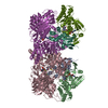
 Assembly


 Mass: 175.820 Da / Num. of mol.: 4 / Source method: obtained synthetically / Formula: Fe2S2
 Mass: 40.078 Da / Num. of mol.: 2 / Source method: obtained synthetically / Formula: Ca
 Mass: 785.550 Da / Num. of mol.: 2 / Source method: obtained synthetically / Formula: C27H33N9O15P2 / Comment: FAD*YM
 Mass: 665.441 Da / Num. of mol.: 2 / Source method: obtained synthetically / Formula: C21H29N7O14P2
 Mass: 61.017 Da / Num. of mol.: 2 / Source method: obtained synthetically / Formula: CHO3 / Comment: pH buffer*YM
 Mass: 92.094 Da / Num. of mol.: 3 / Source method: obtained synthetically / Formula: C3H8O3
 Mass: 395.352 Da / Num. of mol.: 2 / Source method: obtained synthetically / Formula: C10H14N5O6PS2
 Mass: 161.012 Da / Num. of mol.: 2 / Source method: obtained synthetically / Formula: HMoO2S
 Mass: 168.110 Da / Num. of mol.: 2 / Source method: obtained synthetically / Formula: C5H4N4O3
 Sample preparation
 / Beamline: AR-NW12A / Wavelength: 1 Å
 Processing