 Movie
Movie Controller

[English] 日本語
 Yorodumi
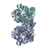
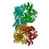
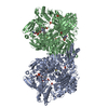
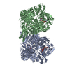
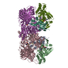
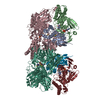
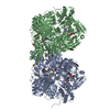
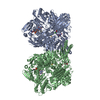
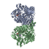
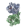
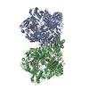
- PDB-2e1q: Crystal Structure of Human Xanthine Oxidoreductase mutant, Glu803Val -
+ Open data

- Basic information
| Entry | Database: PDB / ID: 2e1q | ||||||
|---|---|---|---|---|---|---|---|
| Title | Crystal Structure of Human Xanthine Oxidoreductase mutant, Glu803Val | ||||||
 Components Components | Xanthine dehydrogenase/oxidase | ||||||
 Keywords Keywords | OXIDOREDUCTASE / xanthine oxidase / molybdenum cofactor / FAD | ||||||
| Function / homology |  Function and homology information Function and homology informationhypoxanthine catabolic process / hypoxanthine dehydrogenase activity / hypoxanthine oxidase activity / guanine catabolic process / deoxyguanosine catabolic process / xanthine dehydrogenase / xanthine oxidase / xanthine oxidase activity / xanthine catabolic process / xanthine dehydrogenase activity ...hypoxanthine catabolic process / hypoxanthine dehydrogenase activity / hypoxanthine oxidase activity / guanine catabolic process / deoxyguanosine catabolic process / xanthine dehydrogenase / xanthine oxidase / xanthine oxidase activity / xanthine catabolic process / xanthine dehydrogenase activity / GMP catabolic process / deoxyinosine catabolic process / regulation of epithelial cell differentiation / deoxyadenosine catabolic process / dAMP catabolic process / inosine catabolic process / : / dGMP catabolic process / AMP catabolic process / Butyrophilin (BTN) family interactions / adenosine catabolic process / IMP catabolic process / allantoin metabolic process / Purine catabolism / molybdopterin cofactor binding / Azathioprine ADME / iron-sulfur cluster assembly / lactation / FAD binding / sarcoplasmic reticulum / 2 iron, 2 sulfur cluster binding / flavin adenine dinucleotide binding / peroxisome / iron ion binding / protein homodimerization activity / : / cytosol Similarity search - Function | ||||||
| Biological species |  Homo sapiens (human) Homo sapiens (human) | ||||||
| Method |  X-RAY DIFFRACTION / SYNCHROTRON / MOLECULAR REPLACEMENT / Resolution: 2.6 Å X-RAY DIFFRACTION / SYNCHROTRON / MOLECULAR REPLACEMENT / Resolution: 2.6 Å | ||||||
 Authors Authors | Yamaguchi, Y. / Matsumura, T. / Ichida, K. / Okamoto, K. / Nishino, T. | ||||||
 Citation Citation | Journal: J.Biochem.(Tokyo) / Year: 2007 Title: Human xanthine oxidase changes its substrate specificity to aldehyde oxidase type upon mutation of amino acid residues in the active site: roles of active site residues in binding and ...Title: Human xanthine oxidase changes its substrate specificity to aldehyde oxidase type upon mutation of amino acid residues in the active site: roles of active site residues in binding and activation of purine substrate Authors: Yamaguchi, Y. / Matsumura, T. / Ichida, K. / Okamoto, K. / Nishino, T. | ||||||
| History |
|
- Structure visualization
| Structure viewer | Molecule: MolmilJmol/JSmol |
|---|
- Downloads & links
-Download
| PDBx/mmCIF format | 2e1q.cif.gz | 1022.5 KB | Display | PDBx/mmCIF format |
|---|---|---|---|---|
| PDB format | pdb2e1q.ent.gz | 831.3 KB | Display | PDB format |
| PDBx/mmJSON format | 2e1q.json.gz | Tree view | PDBx/mmJSON format | |
| Others |  Other downloads Other downloads |
-Validation report
| Arichive directory | https://data.pdbj.org/pub/pdb/validation_reports/e1/2e1qftp://data.pdbj.org/pub/pdb/validation_reports/e1/2e1q | HTTPS FTP |
|---|
-Related structure data
| Related structure data | 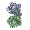 1fo4S S: Starting model for refinement |
|---|---|
| Similar structure data |


-Links
 PDBj
PDBj


- Assembly
| Deposited unit | 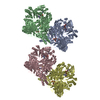
| ||||||||
|---|---|---|---|---|---|---|---|---|---|
| 1 | 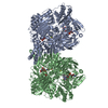
| ||||||||
| 2 | 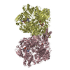
| ||||||||
| Unit cell |
| ||||||||
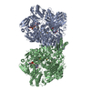
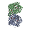
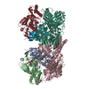
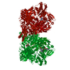
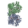
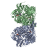
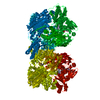
| Details | The biological assembly is a homodimer. |
-Components
-Protein , 1 types, 4 molecules ABCD
| #1: Protein | Mass: 146571.797 Da / Num. of mol.: 4 / Mutation: E803V Source method: isolated from a genetically manipulated source Source: (gene. exp.) Homo sapiens (human) / Plasmid: pTrc99A / Production host:  References: UniProt: P47989, xanthine dehydrogenase, xanthine oxidase |
|---|
-Non-polymers , 8 types, 985 molecules 


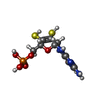
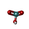

| #2: Chemical | ChemComp-BCT /  Mass: 61.017 Da / Num. of mol.: 4 / Source method: obtained synthetically / Formula: CHO3 / Comment: pH buffer*YM Mass: 61.017 Da / Num. of mol.: 4 / Source method: obtained synthetically / Formula: CHO3 / Comment: pH buffer*YM#3: Chemical | ChemComp-CA /  Mass: 40.078 Da / Num. of mol.: 8 / Source method: obtained synthetically / Formula: Ca Mass: 40.078 Da / Num. of mol.: 8 / Source method: obtained synthetically / Formula: Ca#4: Chemical | ChemComp-FES /  Mass: 175.820 Da / Num. of mol.: 8 / Source method: obtained synthetically / Formula: Fe2S2 Mass: 175.820 Da / Num. of mol.: 8 / Source method: obtained synthetically / Formula: Fe2S2#5: Chemical | ChemComp-FAD /  Mass: 785.550 Da / Num. of mol.: 4 / Source method: obtained synthetically / Formula: C27H33N9O15P2 / Comment: FAD*YM Mass: 785.550 Da / Num. of mol.: 4 / Source method: obtained synthetically / Formula: C27H33N9O15P2 / Comment: FAD*YM#6: Chemical | ChemComp-MTE /  Mass: 395.352 Da / Num. of mol.: 4 / Source method: obtained synthetically / Formula: C10H14N5O6PS2 Mass: 395.352 Da / Num. of mol.: 4 / Source method: obtained synthetically / Formula: C10H14N5O6PS2#7: Chemical | ChemComp-MOM /  Mass: 144.946 Da / Num. of mol.: 4 / Source method: obtained synthetically / Formula: HMoO3 Mass: 144.946 Da / Num. of mol.: 4 / Source method: obtained synthetically / Formula: HMoO3#8: Chemical | ChemComp-SAL /  Mass: 138.121 Da / Num. of mol.: 4 / Source method: obtained synthetically / Formula: C7H6O3 Mass: 138.121 Da / Num. of mol.: 4 / Source method: obtained synthetically / Formula: C7H6O3#9: Water | ChemComp-HOH / | Mass: 18.015 Da / Num. of mol.: 949 / Source method: isolated from a natural source / Formula: H2O |
|---|
-Experimental details
-Experiment
| Experiment | Method: X-RAY DIFFRACTION / Number of used crystals: 1 |
|---|
- Sample preparation
| Crystal | Density Matthews: 2.85 Å3/Da / Density % sol: 56.88 % |
|---|---|
| Crystal grow | Temperature: 293 K / Method: vapor diffusion / pH: 5 Details: 5% PEG 8000, 0.05M sodium citrate, pH 5.0, VAPOR DIFFUSION, temperature 293K |
-Data collection
| Diffraction source | Source: SYNCHROTRON / Site: Photon Factory  / Beamline: BL-6A / Wavelength: 1 Å / Beamline: BL-6A / Wavelength: 1 Å |
|---|---|
| Detector | Type: ADSC QUANTUM 4 / Detector: CCD |
| Radiation | Protocol: SINGLE WAVELENGTH / Monochromatic (M) / Laue (L): M / Scattering type: x-ray |
| Radiation wavelength | Wavelength: 1 Å / Relative weight: 1 |
| Reflection | Resolution: 2.6→50 Å / Num. all: 202081 / Num. obs: 192029 / % possible obs: 95 % / Rsym value: 0.085 |
- Processing
| Software |
| ||||||||||||||||||||
|---|---|---|---|---|---|---|---|---|---|---|---|---|---|---|---|---|---|---|---|---|---|
| Refinement | Method to determine structure: MOLECULAR REPLACEMENT Starting model: PDB ENTRY 1FO4 Resolution: 2.6→50 Å
| ||||||||||||||||||||
| Refinement step | Cycle: LAST / Resolution: 2.6→50 Å
| ||||||||||||||||||||
| Refine LS restraints |
|
