 Movie
Movie Controller


+ Open data

- Basic information
| Entry | Database: PDB / ID: 3eub | ||||||
|---|---|---|---|---|---|---|---|
| Title | Crystal Structure of Desulfo-Xanthine Oxidase with Xanthine | ||||||
 Components Components | (Xanthine dehydrogenase/oxidase) x 3 | ||||||
 Keywords Keywords | OXIDOREDUCTASE / enzyme catalysis / desulfo / substrate orientation / xanthine / FAD / Flavoprotein / Iron / Iron-sulfur / Metal-binding / Molybdenum / NAD / Peroxisome | ||||||
| Function / homology |  Function and homology information Function and homology informationxanthine dehydrogenase complex / xanthine dehydrogenase / xanthine oxidase / xanthine oxidase activity / xanthine catabolic process / xanthine dehydrogenase activity / molybdenum ion binding / molybdopterin cofactor binding / FAD binding / 2 iron, 2 sulfur cluster binding ...xanthine dehydrogenase complex / xanthine dehydrogenase / xanthine oxidase / xanthine oxidase activity / xanthine catabolic process / xanthine dehydrogenase activity / molybdenum ion binding / molybdopterin cofactor binding / FAD binding / 2 iron, 2 sulfur cluster binding / flavin adenine dinucleotide binding / peroxisome / iron ion binding / protein homodimerization activity / : Similarity search - Function | ||||||
| Biological species |  | ||||||
| Method |  X-RAY DIFFRACTION / SYNCHROTRON / MOLECULAR REPLACEMENT / molecular replacement / Resolution: 2.6 Å X-RAY DIFFRACTION / SYNCHROTRON / MOLECULAR REPLACEMENT / molecular replacement / Resolution: 2.6 Å | ||||||
 Authors Authors | Pauff, J.M. / Cao, H. / Hille, R. | ||||||
 Citation Citation | Journal: J.Biol.Chem. / Year: 2009 Title: Substrate Orientation and Catalysis at the Molybdenum Site in Xanthine Oxidase: CRYSTAL STRUCTURES IN COMPLEX WITH XANTHINE AND LUMAZINE. Authors: Pauff, J.M. / Cao, H. / Hille, R. | ||||||
| History |
|
- Structure visualization
| Structure viewer | Molecule: MolmilJmol/JSmol |
|---|
- Downloads & links
-Download
| PDBx/mmCIF format | 3eub.cif.gz | 941.6 KB | Display | PDBx/mmCIF format |
|---|---|---|---|---|
| PDB format | pdb3eub.ent.gz | 752.8 KB | Display | PDB format |
| PDBx/mmJSON format | 3eub.json.gz | Tree view | PDBx/mmJSON format | |
| Others |  Other downloads Other downloads |
-Validation report
| Arichive directory | https://data.pdbj.org/pub/pdb/validation_reports/eu/3eubftp://data.pdbj.org/pub/pdb/validation_reports/eu/3eub | HTTPS FTP |
|---|
-Related structure data
| Related structure data | 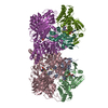 3etrC  1fiqS S: Starting model for refinement C: citing same article ( |
|---|---|
| Similar structure data |

-Links
 PDBj
PDBj


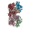
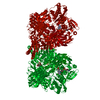
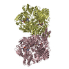
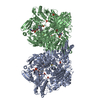
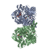
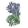
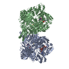
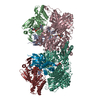
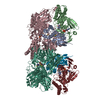
- Assembly
| Deposited unit | 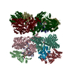
| ||||||||||||||||||||||||||||||||||||||||||||||||||||||||||||||||||||||||||||||||||||||||||||||||||||||||||
|---|---|---|---|---|---|---|---|---|---|---|---|---|---|---|---|---|---|---|---|---|---|---|---|---|---|---|---|---|---|---|---|---|---|---|---|---|---|---|---|---|---|---|---|---|---|---|---|---|---|---|---|---|---|---|---|---|---|---|---|---|---|---|---|---|---|---|---|---|---|---|---|---|---|---|---|---|---|---|---|---|---|---|---|---|---|---|---|---|---|---|---|---|---|---|---|---|---|---|---|---|---|---|---|---|---|---|---|
| 1 | 
| ||||||||||||||||||||||||||||||||||||||||||||||||||||||||||||||||||||||||||||||||||||||||||||||||||||||||||
| 2 | 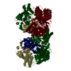
| ||||||||||||||||||||||||||||||||||||||||||||||||||||||||||||||||||||||||||||||||||||||||||||||||||||||||||
| Unit cell |
| ||||||||||||||||||||||||||||||||||||||||||||||||||||||||||||||||||||||||||||||||||||||||||||||||||||||||||
| Noncrystallographic symmetry (NCS) | NCS domain:
NCS domain segments: Dom-ID: 1 / Refine code: 1
NCS ensembles :
|
-Components
-Protein , 3 types, 12 molecules AJS2BKT3CLU4
| #1: Protein | Mass: 18134.078 Da / Num. of mol.: 4 / Fragment: 2Fe-2S ferredoxin-type domain, residues 1-165 / Source method: isolated from a natural source / Source: (natural) References: UniProt: P80457, xanthine dehydrogenase, xanthine oxidase #2: Protein | Mass: 34024.590 Da / Num. of mol.: 4 / Fragment: AD-binding PCMH-type domain, residues 224-528 / Source method: isolated from a natural source / Source: (natural) References: UniProt: P80457, xanthine dehydrogenase, xanthine oxidase #3: Protein | Mass: 83835.273 Da / Num. of mol.: 4 / Fragment: residues 571-1332 / Source method: isolated from a natural source / Source: (natural) References: UniProt: P80457, xanthine dehydrogenase, xanthine oxidase |
|---|
-Non-polymers , 5 types, 24 molecules 

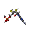
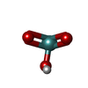
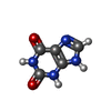
| #4: Chemical | ChemComp-FES /  Mass: 175.820 Da / Num. of mol.: 8 / Source method: obtained synthetically / Formula: Fe2S2 Mass: 175.820 Da / Num. of mol.: 8 / Source method: obtained synthetically / Formula: Fe2S2#5: Chemical | ChemComp-FAD /  Mass: 785.550 Da / Num. of mol.: 4 / Source method: obtained synthetically / Formula: C27H33N9O15P2 / Comment: FAD*YM Mass: 785.550 Da / Num. of mol.: 4 / Source method: obtained synthetically / Formula: C27H33N9O15P2 / Comment: FAD*YM#6: Chemical | ChemComp-MTE /  Mass: 395.352 Da / Num. of mol.: 4 / Source method: obtained synthetically / Formula: C10H14N5O6PS2 Mass: 395.352 Da / Num. of mol.: 4 / Source method: obtained synthetically / Formula: C10H14N5O6PS2#7: Chemical | ChemComp-MOM /  Mass: 144.946 Da / Num. of mol.: 4 / Source method: obtained synthetically / Formula: HMoO3 Mass: 144.946 Da / Num. of mol.: 4 / Source method: obtained synthetically / Formula: HMoO3#8: Chemical | ChemComp-XAN /  Mass: 152.111 Da / Num. of mol.: 4 / Source method: obtained synthetically / Formula: C5H4N4O2 Mass: 152.111 Da / Num. of mol.: 4 / Source method: obtained synthetically / Formula: C5H4N4O2 |
|---|
-Experimental details
-Experiment
| Experiment | Method: X-RAY DIFFRACTION / Number of used crystals: 1 |
|---|
- Sample preparation
| Crystal | Density Matthews: 2.54 Å3/Da / Density % sol: 51.52 % |
|---|---|
| Crystal grow | Temperature: 298 K / Method: batch / pH: 7.2 / Details: PEG 8000, pH 7.2, batch, temperature 298K |
-Data collection
| Diffraction | Mean temperature: 100 K |
|---|---|
| Diffraction source | Source: SYNCHROTRON / Site: APS  / Beamline: 31-ID / Wavelength: 0.9793 Å / Beamline: 31-ID / Wavelength: 0.9793 Å |
| Detector | Type: MAR CCD 165 mm / Detector: CCD / Date: Mar 20, 2008 |
| Radiation | Protocol: SINGLE WAVELENGTH / Monochromatic (M) / Laue (L): M / Scattering type: x-ray |
| Radiation wavelength | Wavelength: 0.9793 Å / Relative weight: 1 |
| Reflection | Resolution: 2.6→141.42 Å / Num. obs: 119504 |
| Reflection shell | Resolution: 2.6→2.668 Å / Num. unique all: 5403 |
-Phasing
| Phasing | Method: molecular replacement | |||||||||
|---|---|---|---|---|---|---|---|---|---|---|
| Phasing MR |
|
- Processing
| Software |
| ||||||||||||||||||||||||||||||||||||||||||||||||||||||||||||||||||||||||||||||||||||||||||
|---|---|---|---|---|---|---|---|---|---|---|---|---|---|---|---|---|---|---|---|---|---|---|---|---|---|---|---|---|---|---|---|---|---|---|---|---|---|---|---|---|---|---|---|---|---|---|---|---|---|---|---|---|---|---|---|---|---|---|---|---|---|---|---|---|---|---|---|---|---|---|---|---|---|---|---|---|---|---|---|---|---|---|---|---|---|---|---|---|---|---|---|
| Refinement | Method to determine structure: MOLECULAR REPLACEMENT Starting model: PDB entry 1FIQ Resolution: 2.6→33.08 Å / Cor.coef. Fo:Fc: 0.889 / Cor.coef. Fo:Fc free: 0.826 / WRfactor Rfree: 0.308 / WRfactor Rwork: 0.246 / Occupancy max: 1 / Occupancy min: 1 / FOM work R set: 0.823 / SU B: 12.029 / SU ML: 0.263 / SU Rfree: 0.457 / Cross valid method: THROUGHOUT / σ(F): 0 / ESU R Free: 0.457 / Stereochemistry target values: MAXIMUM LIKELIHOOD Details: HYDROGENS HAVE BEEN ADDED IN THE RIDING POSITIONS, MOM IS IN THE DESULFO FORM WITH AN ADDITIONAL OXYGEN IN PLACE OF THE SULFUR IN THE COORDINATION SPHERE
| ||||||||||||||||||||||||||||||||||||||||||||||||||||||||||||||||||||||||||||||||||||||||||
| Solvent computation | Ion probe radii: 0.8 Å / Shrinkage radii: 0.8 Å / VDW probe radii: 1.2 Å / Solvent model: MASK | ||||||||||||||||||||||||||||||||||||||||||||||||||||||||||||||||||||||||||||||||||||||||||
| Displacement parameters | Biso max: 50.14 Å2 / Biso mean: 13.895 Å2 / Biso min: 2 Å2
| ||||||||||||||||||||||||||||||||||||||||||||||||||||||||||||||||||||||||||||||||||||||||||
| Refinement step | Cycle: LAST / Resolution: 2.6→33.08 Å
| ||||||||||||||||||||||||||||||||||||||||||||||||||||||||||||||||||||||||||||||||||||||||||
| Refine LS restraints |
| ||||||||||||||||||||||||||||||||||||||||||||||||||||||||||||||||||||||||||||||||||||||||||
| Refine LS restraints NCS | Dom-ID: 1 / Refine-ID: X-RAY DIFFRACTION
| ||||||||||||||||||||||||||||||||||||||||||||||||||||||||||||||||||||||||||||||||||||||||||
| LS refinement shell | Resolution: 2.6→2.668 Å / Total num. of bins used: 20
|
