 Movie
Movie Controller

[English] 日本語
 Yorodumi
- PDB-3ueo: Crystal structure of TopBP1 BRCT4/5 domains in complex with a pho... -
+ Open data

- Basic information
| Entry | Database: PDB / ID: 3ueo | ||||||
|---|---|---|---|---|---|---|---|
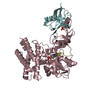
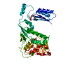
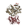
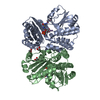
| Title | Crystal structure of TopBP1 BRCT4/5 domains in complex with a phospho-peptide | ||||||
 Components Components |
| ||||||
 Keywords Keywords | PEPTIDE BINDING PROTEIN / BRCT domain / phospho-peptide binding | ||||||
| Function / homology |  Function and homology information Function and homology informationbroken chromosome clustering / BRCA1-B complex / phosphorylation-dependent protein binding / homologous recombination / DNA replication checkpoint signaling / double-strand break repair via classical nonhomologous end joining / double-strand break repair via alternative nonhomologous end joining / mitotic DNA replication checkpoint signaling / protein localization to site of double-strand break / chromatin-protein adaptor activity ...broken chromosome clustering / BRCA1-B complex / phosphorylation-dependent protein binding / homologous recombination / DNA replication checkpoint signaling / double-strand break repair via classical nonhomologous end joining / double-strand break repair via alternative nonhomologous end joining / mitotic DNA replication checkpoint signaling / protein localization to site of double-strand break / chromatin-protein adaptor activity / mitotic intra-S DNA damage checkpoint signaling / HDR through Single Strand Annealing (SSA) / DNA metabolic process / response to ionizing radiation / mitotic G2 DNA damage checkpoint signaling / Impaired BRCA2 binding to RAD51 / chromosome organization / Presynaptic phase of homologous DNA pairing and strand exchange / DNA replication initiation / histone reader activity / SUMOylation of DNA damage response and repair proteins / site of DNA damage / DNA damage checkpoint signaling / male germ cell nucleus / condensed nuclear chromosome / protein serine/threonine kinase activator activity / TP53 Regulates Transcription of DNA Repair Genes / Nonhomologous End-Joining (NHEJ) / PML body / G2/M DNA damage checkpoint / double-strand break repair via homologous recombination / spindle pole / actin cytoskeleton / chromosome / Recruitment and ATM-mediated phosphorylation of repair and signaling proteins at DNA double strand breaks / site of double-strand break / Processing of DNA double-strand break ends / Regulation of TP53 Activity through Phosphorylation / nuclear body / focal adhesion / DNA repair / centrosome / DNA damage response / DNA binding / nucleoplasm / identical protein binding / nucleus / plasma membrane Similarity search - Function | ||||||
| Biological species |  Homo sapiens (human) Homo sapiens (human) | ||||||
| Method |  X-RAY DIFFRACTION / SYNCHROTRON / MOLECULAR REPLACEMENT / molecular replacement / Resolution: 2.6 Å X-RAY DIFFRACTION / SYNCHROTRON / MOLECULAR REPLACEMENT / molecular replacement / Resolution: 2.6 Å | ||||||
 Authors Authors | Leung, C.C. / Glover, J.N.M. | ||||||
 Citation Citation | Journal: Structure / Year: 2013 Title: Structural insights into recognition of MDC1 by TopBP1 in DNA replication checkpoint control. Authors: Leung, C.C. / Sun, L. / Gong, Z. / Burkat, M. / Edwards, R. / Assmus, M. / Chen, J. / Glover, J.N. | ||||||
| History |
|
- Structure visualization
| Structure viewer | Molecule: MolmilJmol/JSmol |
|---|
- Downloads & links
-Download
| PDBx/mmCIF format | 3ueo.cif.gz | 306 KB | Display | PDBx/mmCIF format |
|---|---|---|---|---|
| PDB format | pdb3ueo.ent.gz | 249.6 KB | Display | PDB format |
| PDBx/mmJSON format | 3ueo.json.gz | Tree view | PDBx/mmJSON format | |
| Others |  Other downloads Other downloads |
-Validation report
| Arichive directory | https://data.pdbj.org/pub/pdb/validation_reports/ue/3ueoftp://data.pdbj.org/pub/pdb/validation_reports/ue/3ueo | HTTPS FTP |
|---|
-Related structure data


-Links
 PDBj
PDBj


- Assembly
| Deposited unit | 
| ||||||||||||||||||||||||||||||||||||||||||||||||||||||||||||
|---|---|---|---|---|---|---|---|---|---|---|---|---|---|---|---|---|---|---|---|---|---|---|---|---|---|---|---|---|---|---|---|---|---|---|---|---|---|---|---|---|---|---|---|---|---|---|---|---|---|---|---|---|---|---|---|---|---|---|---|---|---|
| 1 | 
| ||||||||||||||||||||||||||||||||||||||||||||||||||||||||||||
| 2 | 
| ||||||||||||||||||||||||||||||||||||||||||||||||||||||||||||
| Unit cell |
| ||||||||||||||||||||||||||||||||||||||||||||||||||||||||||||
| Noncrystallographic symmetry (NCS) | NCS domain:
NCS domain segments:
NCS ensembles :
NCS oper:
|
-Components
| #1: Protein | Mass: 22238.371 Da / Num. of mol.: 4 / Fragment: BRCT domain Source method: isolated from a genetically manipulated source Source: (gene. exp.) Homo sapiens (human) / Gene: KIAA0259, TOPBP1 / Plasmid: pGEX / Production host:  #2: Protein/peptide | Mass: 1515.275 Da / Num. of mol.: 2 / Source method: obtained synthetically / Details: This represents a consensus sequence in humans / References: UniProt: Q14676*PLUS #3: Water | ChemComp-HOH / |  Mass: 18.015 Da / Num. of mol.: 257 / Source method: isolated from a natural source / Formula: H2O Mass: 18.015 Da / Num. of mol.: 257 / Source method: isolated from a natural source / Formula: H2OHas protein modification | Y | |
|---|
-Experimental details
-Experiment
| Experiment | Method: X-RAY DIFFRACTION / Number of used crystals: 1 |
|---|
- Sample preparation
| Crystal | Density Matthews: 2.55 Å3/Da / Density % sol: 51.77 % |
|---|---|
| Crystal grow | Temperature: 298 K / Method: vapor diffusion, hanging drop / pH: 5.5 Details: PEG 10000, Ammonium acetate, bis-Tris, pH 5.5, VAPOR DIFFUSION, HANGING DROP, temperature 298K |
-Data collection
| Diffraction | Mean temperature: 298 K | ||||||||||||||||||||||||||||||||||||||||||||||||||||||||||||||||||||||
|---|---|---|---|---|---|---|---|---|---|---|---|---|---|---|---|---|---|---|---|---|---|---|---|---|---|---|---|---|---|---|---|---|---|---|---|---|---|---|---|---|---|---|---|---|---|---|---|---|---|---|---|---|---|---|---|---|---|---|---|---|---|---|---|---|---|---|---|---|---|---|---|
| Diffraction source | Source: SYNCHROTRON / Site: CLSI  / Beamline: 08ID-1 / Wavelength: 0.97946 Å / Beamline: 08ID-1 / Wavelength: 0.97946 Å | ||||||||||||||||||||||||||||||||||||||||||||||||||||||||||||||||||||||
| Detector | Type: MARMOSAIC 325 mm CCD / Detector: CCD / Date: Feb 6, 2011 | ||||||||||||||||||||||||||||||||||||||||||||||||||||||||||||||||||||||
| Radiation | Protocol: SINGLE WAVELENGTH / Monochromatic (M) / Laue (L): M / Scattering type: x-ray | ||||||||||||||||||||||||||||||||||||||||||||||||||||||||||||||||||||||
| Radiation wavelength | Wavelength: 0.97946 Å / Relative weight: 1 | ||||||||||||||||||||||||||||||||||||||||||||||||||||||||||||||||||||||
| Reflection | Resolution: 2.6→34.6 Å / Num. all: 26653 / Num. obs: 26653 / % possible obs: 95.2 % / Observed criterion σ(I): -3 / Biso Wilson estimate: 47.8 Å2 / Rmerge(I) obs: 0.059 / Net I/σ(I): 11.89 | ||||||||||||||||||||||||||||||||||||||||||||||||||||||||||||||||||||||
| Reflection shell | Diffraction-ID: 1
|
-Phasing
| Phasing | Method: molecular replacement | |||||||||
|---|---|---|---|---|---|---|---|---|---|---|
| Phasing MR | Rfactor: 43.49 / Model details: Phaser MODE: MR_AUTO
|
- Processing
| Software |
| |||||||||||||||||||||||||||||||||||||||||||||||||||||||||||||||||||||||||||||||||||||||||||||||||||||||||||||||||||||||||||||||||||||||||||||||||||||||||||||||||||||||||||||||
|---|---|---|---|---|---|---|---|---|---|---|---|---|---|---|---|---|---|---|---|---|---|---|---|---|---|---|---|---|---|---|---|---|---|---|---|---|---|---|---|---|---|---|---|---|---|---|---|---|---|---|---|---|---|---|---|---|---|---|---|---|---|---|---|---|---|---|---|---|---|---|---|---|---|---|---|---|---|---|---|---|---|---|---|---|---|---|---|---|---|---|---|---|---|---|---|---|---|---|---|---|---|---|---|---|---|---|---|---|---|---|---|---|---|---|---|---|---|---|---|---|---|---|---|---|---|---|---|---|---|---|---|---|---|---|---|---|---|---|---|---|---|---|---|---|---|---|---|---|---|---|---|---|---|---|---|---|---|---|---|---|---|---|---|---|---|---|---|---|---|---|---|---|---|---|---|---|
| Refinement | Method to determine structure: MOLECULAR REPLACEMENT / Resolution: 2.6→34.566 Å / Occupancy max: 1 / Occupancy min: 0.5 / FOM work R set: 0.8071 / SU ML: 0.69 / σ(F): 1.99 / Phase error: 26.58 / Stereochemistry target values: ML
| |||||||||||||||||||||||||||||||||||||||||||||||||||||||||||||||||||||||||||||||||||||||||||||||||||||||||||||||||||||||||||||||||||||||||||||||||||||||||||||||||||||||||||||||
| Solvent computation | Shrinkage radii: 0.9 Å / VDW probe radii: 1.11 Å / Solvent model: FLAT BULK SOLVENT MODEL / Bsol: 39.676 Å2 / ksol: 0.333 e/Å3 | |||||||||||||||||||||||||||||||||||||||||||||||||||||||||||||||||||||||||||||||||||||||||||||||||||||||||||||||||||||||||||||||||||||||||||||||||||||||||||||||||||||||||||||||
| Displacement parameters | Biso max: 166.19 Å2 / Biso mean: 44.2076 Å2 / Biso min: 4.6 Å2
| |||||||||||||||||||||||||||||||||||||||||||||||||||||||||||||||||||||||||||||||||||||||||||||||||||||||||||||||||||||||||||||||||||||||||||||||||||||||||||||||||||||||||||||||
| Refinement step | Cycle: LAST / Resolution: 2.6→34.566 Å
| |||||||||||||||||||||||||||||||||||||||||||||||||||||||||||||||||||||||||||||||||||||||||||||||||||||||||||||||||||||||||||||||||||||||||||||||||||||||||||||||||||||||||||||||
| Refine LS restraints |
| |||||||||||||||||||||||||||||||||||||||||||||||||||||||||||||||||||||||||||||||||||||||||||||||||||||||||||||||||||||||||||||||||||||||||||||||||||||||||||||||||||||||||||||||
| Refine LS restraints NCS |
| |||||||||||||||||||||||||||||||||||||||||||||||||||||||||||||||||||||||||||||||||||||||||||||||||||||||||||||||||||||||||||||||||||||||||||||||||||||||||||||||||||||||||||||||
| LS refinement shell | Refine-ID: X-RAY DIFFRACTION / Total num. of bins used: 9
| |||||||||||||||||||||||||||||||||||||||||||||||||||||||||||||||||||||||||||||||||||||||||||||||||||||||||||||||||||||||||||||||||||||||||||||||||||||||||||||||||||||||||||||||
| Refinement TLS params. | Method: refined / Refine-ID: X-RAY DIFFRACTION
| |||||||||||||||||||||||||||||||||||||||||||||||||||||||||||||||||||||||||||||||||||||||||||||||||||||||||||||||||||||||||||||||||||||||||||||||||||||||||||||||||||||||||||||||
| Refinement TLS group |
|
