 Movie
Movie Controller

[English] 日本語
 Yorodumi
- PDB-2yk6: Structure of Neisseria LOS-specific sialyltransferase (NST), in c... -
+ Open data

- Basic information
| Entry | Database: PDB / ID: 2yk6 | ||||||
|---|---|---|---|---|---|---|---|
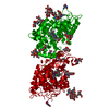
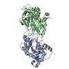
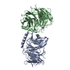
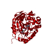
| Title | Structure of Neisseria LOS-specific sialyltransferase (NST), in complex with CDP. | ||||||
 Components Components | CMP-N-ACETYLNEURAMINATE-BETA-GALACTOSAMIDE-ALPHA-2,3-SIALYLTRANSFERASE | ||||||
 Keywords Keywords | TRANSFERASE / LIPOOLIGOSACCHARIDE SIALYLTRANSFERASE | ||||||
| Function / homology |  Function and homology information Function and homology informationN-acetyllactosaminide alpha-2,3-sialyltransferase / N-acetyllactosaminide alpha-2,3-sialyltransferase activity / lipopolysaccharide biosynthetic process / cell outer membrane Similarity search - Function | ||||||
| Biological species |  NEISSERIA MENINGITIDIS SEROGROUP B (bacteria) NEISSERIA MENINGITIDIS SEROGROUP B (bacteria) | ||||||
| Method |  X-RAY DIFFRACTION / MOLECULAR REPLACEMENT / Resolution: 2.83 Å X-RAY DIFFRACTION / MOLECULAR REPLACEMENT / Resolution: 2.83 Å | ||||||
 Authors Authors | Lin, L.Y.C. / Rakic, B. / Chiu, C.P.C. / Lameignere, E. / Wakarchuk, W.W. / Withers, S.G. / Strynadka, N.C.J. | ||||||
 Citation Citation | Journal: J.Biol.Chem. / Year: 2011 Title: Structure and Mechanism of the Lipooligosaccharide Sialyltransferase from Neisseria Meningitidis Authors: Lin, L.Y.C. / Rakic, B. / Chiu, C.P.C. / Lameignere, E. / Wakarchuk, W.W. / Withers, S.G. / Strynadka, N.C.J. | ||||||
| History |
|
- Structure visualization
| Structure viewer | Molecule: MolmilJmol/JSmol |
|---|
- Downloads & links
-Download
| PDBx/mmCIF format | 2yk6.cif.gz | 84.3 KB | Display | PDBx/mmCIF format |
|---|---|---|---|---|
| PDB format | pdb2yk6.ent.gz | 62 KB | Display | PDB format |
| PDBx/mmJSON format | 2yk6.json.gz | Tree view | PDBx/mmJSON format | |
| Others |  Other downloads Other downloads |
-Validation report
| Arichive directory | https://data.pdbj.org/pub/pdb/validation_reports/yk/2yk6ftp://data.pdbj.org/pub/pdb/validation_reports/yk/2yk6 | HTTPS FTP |
|---|
-Related structure data
| Related structure data |  2yk4C  2yk5SC  2yk7C C: citing same article ( S: Starting model for refinement |
|---|---|
| Similar structure data |


-Links
 PDBj
PDBj- Assembly
| Deposited unit | 
| ||||||||
|---|---|---|---|---|---|---|---|---|---|
| 1 | 
| ||||||||
| Unit cell |
|
-Components
| #1: Protein | Mass: 37565.203 Da / Num. of mol.: 1 / Fragment: DELTA29NST, RESIDUES 49-370 Source method: isolated from a genetically manipulated source Source: (gene. exp.) NEISSERIA MENINGITIDIS SEROGROUP B (bacteria)Strain: 126E / NRCC4010 / Variant: L1 IMMUNOTYPE / Production host: References: UniProt: P72097, Transferases; Glycosyltransferases; Transferring other glycosyl groups |
|---|---|
| #2: Chemical | ChemComp-CDP /   Mass: 403.176 Da / Num. of mol.: 1 / Source method: obtained synthetically / Formula: C9H15N3O11P2 Mass: 403.176 Da / Num. of mol.: 1 / Source method: obtained synthetically / Formula: C9H15N3O11P2 |
| #3: Chemical | ChemComp-1PE /   Mass: 238.278 Da / Num. of mol.: 1 / Source method: obtained synthetically / Formula: C10H22O6 / Comment: precipitant*YM Mass: 238.278 Da / Num. of mol.: 1 / Source method: obtained synthetically / Formula: C10H22O6 / Comment: precipitant*YM |
| #4: Chemical | ChemComp-SO4 /   Mass: 96.063 Da / Num. of mol.: 1 / Source method: obtained synthetically / Formula: SO4 Mass: 96.063 Da / Num. of mol.: 1 / Source method: obtained synthetically / Formula: SO4 |
| #5: Water | ChemComp-HOH /  Mass: 18.015 Da / Num. of mol.: 81 / Source method: isolated from a natural source / Formula: H2O Mass: 18.015 Da / Num. of mol.: 81 / Source method: isolated from a natural source / Formula: H2O |
| Sequence details | E40D, R102W, G168I, AND K273N ARE NATURAL VARIANTS IN THE STRAIN N. MENINGITID |
-Experimental details
-Experiment
| Experiment | Method: X-RAY DIFFRACTION / Number of used crystals: 1 |
|---|
- Sample preparation
| Crystal | Density Matthews: 2.96 Å3/Da / Density % sol: 58.45 % / Description: NONE |
|---|---|
| Crystal grow | Method: vapor diffusion / pH: 4.4 Details: PROTEIN WAS CRYSTALLIZED BY VAPOR DIFFUSION METHODS USING DROPS OF PROTEIN MIXED WITH AN EQUAL VOLUME OF PRECIPITANT: 100 MM SODIUM ACETATE (PH 4.2 - 4.4) 1.7 M DI-AMMONIUM SULFATE. |
-Data collection
| Diffraction | Mean temperature: 100 K |
|---|---|
| Diffraction source | Source: ROTATING ANODE / Type: RIGAKU MICROMAX-007 HF / Wavelength: 1.54 / Wavelength: 1.54 Å |
| Detector | Type: MARRESEARCH / Detector: IMAGE PLATE / Details: X-RAY MIRRORS OSMIC VARIMAXHR |
| Radiation | Protocol: SINGLE WAVELENGTH / Monochromatic (M) / Laue (L): M / Scattering type: x-ray |
| Radiation wavelength | Wavelength: 1.54 Å / Relative weight: 1 |
| Reflection | Resolution: 2.83→70.71 Å / Num. obs: 11162 / % possible obs: 99.7 % / Observed criterion σ(I): 2 / Redundancy: 6.8 % / Rmerge(I) obs: 0.121 / Net I/σ(I): 15.4 |
| Reflection shell | Resolution: 2.83→2.88 Å / Redundancy: 5.1 % / Rmerge(I) obs: 0.522 / Mean I/σ(I) obs: 2.7 / % possible all: 97.8 |
- Processing
| Software |
| ||||||||||||||||||||||||||||||||||||||||||||||||||||||||||||||||||||||||||||||||||||||||||||||||||||||||||||||||||||||||||||||||||||||||||||||||||||||||||||||||||||||||||||||||||||||
|---|---|---|---|---|---|---|---|---|---|---|---|---|---|---|---|---|---|---|---|---|---|---|---|---|---|---|---|---|---|---|---|---|---|---|---|---|---|---|---|---|---|---|---|---|---|---|---|---|---|---|---|---|---|---|---|---|---|---|---|---|---|---|---|---|---|---|---|---|---|---|---|---|---|---|---|---|---|---|---|---|---|---|---|---|---|---|---|---|---|---|---|---|---|---|---|---|---|---|---|---|---|---|---|---|---|---|---|---|---|---|---|---|---|---|---|---|---|---|---|---|---|---|---|---|---|---|---|---|---|---|---|---|---|---|---|---|---|---|---|---|---|---|---|---|---|---|---|---|---|---|---|---|---|---|---|---|---|---|---|---|---|---|---|---|---|---|---|---|---|---|---|---|---|---|---|---|---|---|---|---|---|---|---|
| Refinement | Method to determine structure: MOLECULAR REPLACEMENT Starting model: PDB ENTRY 2YK5 Resolution: 2.83→70.71 Å / Cor.coef. Fo:Fc: 0.931 / Cor.coef. Fo:Fc free: 0.883 / SU B: 12.501 / SU ML: 0.244 / Cross valid method: THROUGHOUT / ESU R Free: 0.376 / Stereochemistry target values: MAXIMUM LIKELIHOOD / Details: HYDROGENS HAVE BEEN ADDED IN THE RIDING POSITIONS.
| ||||||||||||||||||||||||||||||||||||||||||||||||||||||||||||||||||||||||||||||||||||||||||||||||||||||||||||||||||||||||||||||||||||||||||||||||||||||||||||||||||||||||||||||||||||||
| Solvent computation | Ion probe radii: 0.8 Å / Shrinkage radii: 0.8 Å / VDW probe radii: 1.4 Å / Solvent model: MASK | ||||||||||||||||||||||||||||||||||||||||||||||||||||||||||||||||||||||||||||||||||||||||||||||||||||||||||||||||||||||||||||||||||||||||||||||||||||||||||||||||||||||||||||||||||||||
| Displacement parameters | Biso mean: 27.571 Å2
| ||||||||||||||||||||||||||||||||||||||||||||||||||||||||||||||||||||||||||||||||||||||||||||||||||||||||||||||||||||||||||||||||||||||||||||||||||||||||||||||||||||||||||||||||||||||
| Refinement step | Cycle: LAST / Resolution: 2.83→70.71 Å
| ||||||||||||||||||||||||||||||||||||||||||||||||||||||||||||||||||||||||||||||||||||||||||||||||||||||||||||||||||||||||||||||||||||||||||||||||||||||||||||||||||||||||||||||||||||||
| Refine LS restraints |
|