- PDB-4cuo: Banyan peroxidase with glycosylation -
+
Open data
ID or keywords:
Loading...
-
Basic information
Entry
Database: PDB / ID: 4cuo
Title
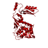
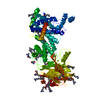
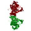
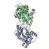
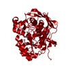
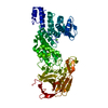
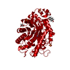
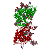
Banyan peroxidase with glycosylation
Components
BANYAN PEROXIDASE
Keywords
OXIDOREDUCTASE / CLASS III / GLYCOSYLATION / SUCCINIMIDE
Function / homology
Function and homology information
lactoperoxidase activity / peroxidase / hydrogen peroxide catabolic process / response to oxidative stress / heme binding / extracellular region / metal ion binding Similarity search - Function
Mass: 18.015 Da / Num. of mol.: 321 / Source method: isolated from a natural source / Formula: H2O
-
Details
Has protein modification
Y
Sequence details
PROTEIN SEQUENCE HAS BEEN DETERMINED FROM THE CRYSTAL STRUCTURE.
-
Experimental details
-
Experiment
Experiment
Method: X-RAY DIFFRACTION / Number of used crystals: 1
-
Sample preparation
Crystal
Density Matthews: 1.8 Å3/Da / Density % sol: 33 % / Description: NONE
Crystal grow
pH: 8 Details: 1 UL PROTEIN (30 MG/ML IN 20 MM TRIS/HCL, PH 8.0) PLUS 1 UL RESERVOIR (1.3 M AMMONIUM SULFATE, 1.0 M LITHIUMSULFATE), CRYOSOLUTION: RESERVOIR INCLUDING 20% GLYCEROL, XE-DERIVATIZATION: 1 MIN ...Details: 1 UL PROTEIN (30 MG/ML IN 20 MM TRIS/HCL, PH 8.0) PLUS 1 UL RESERVOIR (1.3 M AMMONIUM SULFATE, 1.0 M LITHIUMSULFATE), CRYOSOLUTION: RESERVOIR INCLUDING 20% GLYCEROL, XE-DERIVATIZATION: 1 MIN 4 MPA XE FOR CRYSTAL IN CRYOSOLUTION.
Monochromator: MIRROR / Protocol: SINGLE WAVELENGTH / Monochromatic (M) / Laue (L): M / Scattering type: x-ray
Radiation wavelength
ID
Wavelength (Å)
Relative weight
1
1
1
2
1.44163
1
Reflection
Resolution: 1.66→20 Å / Num. obs: 60369 / % possible obs: 99.5 % / Observed criterion σ(I): -3 / Redundancy: 7.4 % / Biso Wilson estimate: 35 Å2 / Rmerge(I) obs: 0.05 / Net I/σ(I): 22.9
Reflection shell
Resolution: 1.66→1.76 Å / Redundancy: 6.9 % / Rmerge(I) obs: 0.99 / Mean I/σ(I) obs: 2.4 / % possible all: 97.7
-
Processing
Software
Name
Version
Classification
REFMAC
5.8.0049
refinement
XDS
datareduction
XDS
datascaling
Auto-Rickshaw
phasing
Refinement
Method to determine structure: SAD Starting model: NONE Resolution: 1.67→63.32 Å / Cor.coef. Fo:Fc: 0.974 / Cor.coef. Fo:Fc free: 0.969 / SU B: 3.888 / SU ML: 0.057 / Cross valid method: THROUGHOUT / ESU R: 0.068 / ESU R Free: 0.07 / Stereochemistry target values: MAXIMUM LIKELIHOOD Details: HYDROGENS HAVE BEEN ADDED IN THE RIDING POSITIONS. HYDROGENS HAVE BEEN USED IF PRESENT IN THE INPUT U VALUES WITH TLS ADDED. NO DNA SEQUENCE WAS AVAILABLE FOR THIS PROTEIN
Rfactor
Num. reflection
% reflection
Selection details
Rfree
0.18029
3049
5.1 %
RANDOM
Rwork
0.15654
-
-
-
obs
0.15776
57238
99.57 %
-
Solvent computation
Ion probe radii: 0.8 Å / Shrinkage radii: 0.6 Å / VDW probe radii: 1.4 Å / Solvent model: BABINET MODEL WITH MASK
 Movie
Movie Controller


 Open data
 Basic information
 Components
 Keywords
 Function and homology information
 FICUS BENGHALENSIS (Indian banyan)
 X-RAY DIFFRACTION /
  Authors
 Citation
 Structure visualization
 Downloads & links
 Other downloads
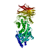

 PDBj
PDBj


 Assembly
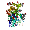

 Type: D-saccharide, beta linking / Mass: 221.208 Da / Num. of mol.: 3


 Mass: 616.487 Da / Num. of mol.: 1 / Source method: obtained synthetically / Formula: C34H32FeN4O4
 Mass: 40.078 Da / Num. of mol.: 2 / Source method: obtained synthetically / Formula: Ca
 Mass: 22.990 Da / Num. of mol.: 1 / Source method: obtained synthetically / Formula: Na
 Mass: 35.453 Da / Num. of mol.: 3 / Source method: obtained synthetically / Formula: Cl
 Mass: 96.063 Da / Num. of mol.: 3 / Source method: obtained synthetically / Formula: SO4
 Mass: 60.009 Da / Num. of mol.: 4 / Source method: obtained synthetically / Formula: CO3
 Sample preparation
 / Beamline: X12 / Wavelength: 1.0, 1.44163
 Processing