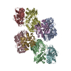
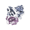
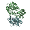
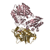
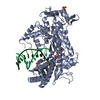
Entry Database : PDB / ID : 6fqbTitle MurT/GatD peptidoglycan amidotransferase complex from Streptococcus pneumoniae R6 Cobyric acid synthase Mur ligase family protein Keywords / / / Function / homology Function Domain/homology Component
/ / / / / / / / / / / / / / / / / / / / / / / / / / / / / / / / / / / / / / / / / / / / / Biological species Streptococcus pneumoniae (bacteria)Method / / / Resolution : 3 Å Authors Morlot, C. / Contreras-Martel, C. / Leisico, F. / Straume, D. / Peters, K. / Hegnar, O.A. / Simon, N. / Villard, A.M. / Breukink, E. / Gravier-Pelletier, C. ...Morlot, C. / Contreras-Martel, C. / Leisico, F. / Straume, D. / Peters, K. / Hegnar, O.A. / Simon, N. / Villard, A.M. / Breukink, E. / Gravier-Pelletier, C. / Le Corre, L. / Vollmer, W. / Pietrancosta, N. / Havarstein, L.S. / Zapun, A. Funding support Organization Grant number Country INSERM-Astra Zeneca
Journal : Nat Commun / Year : 2018Title : Structure of the essential peptidoglycan amidotransferase MurT/GatD complex from Streptococcus pneumoniae.Authors: Morlot, C. / Straume, D. / Peters, K. / Hegnar, O.A. / Simon, N. / Villard, A.M. / Contreras-Martel, C. / Leisico, F. / Breukink, E. / Gravier-Pelletier, C. / Le Corre, L. / Vollmer, W. / ... Authors : Morlot, C. / Straume, D. / Peters, K. / Hegnar, O.A. / Simon, N. / Villard, A.M. / Contreras-Martel, C. / Leisico, F. / Breukink, E. / Gravier-Pelletier, C. / Le Corre, L. / Vollmer, W. / Pietrancosta, N. / Havarstein, L.S. / Zapun, A. History Deposition Feb 13, 2018 Deposition site / Processing site Revision 1.0 Aug 22, 2018 Provider / Type Revision 1.1 Nov 7, 2018 Group / Category Item / _diffrn_source.typeRevision 1.2 Oct 23, 2024 Group Data collection / Database references ... Data collection / Database references / Derived calculations / Refinement description / Structure summary Category chem_comp_atom / chem_comp_bond ... chem_comp_atom / chem_comp_bond / database_2 / pdbx_entry_details / pdbx_modification_feature / struct_conn / struct_ncs_dom_lim Item _database_2.pdbx_DOI / _database_2.pdbx_database_accession ... _database_2.pdbx_DOI / _database_2.pdbx_database_accession / _struct_conn.pdbx_dist_value / _struct_conn.ptnr2_auth_seq_id / _struct_conn.ptnr2_label_seq_id / _struct_ncs_dom_lim.beg_auth_comp_id / _struct_ncs_dom_lim.beg_label_asym_id / _struct_ncs_dom_lim.beg_label_comp_id / _struct_ncs_dom_lim.beg_label_seq_id / _struct_ncs_dom_lim.end_auth_comp_id / _struct_ncs_dom_lim.end_label_asym_id / _struct_ncs_dom_lim.end_label_comp_id / _struct_ncs_dom_lim.end_label_seq_id
Show all Show less
 Movie
Movie Controller

 Yorodumi
 Open data
 Basic information
 Components
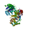
 Keywords
 Function and homology information
 Streptococcus pneumoniae (bacteria)
 X-RAY DIFFRACTION /
  Authors
 France, 1items
  Citation
 Structure visualization
 Downloads & links
 Other downloads


 PDBj
PDBj

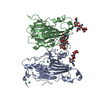
 Assembly