 Movie
Movie Controller

[English] 日本語
 Yorodumi
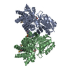
- PDB-3uhj: Crystal structure of a probable glycerol dehydrogenase from Sinor... -
+ Open data

- Basic information
| Entry | Database: PDB / ID: 3uhj | ||||||
|---|---|---|---|---|---|---|---|
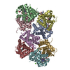
| Title | Crystal structure of a probable glycerol dehydrogenase from Sinorhizobium meliloti 1021 | ||||||
 Components Components | Probable glycerol dehydrogenase | ||||||
 Keywords Keywords | OXIDOREDUCTASE / Structural Genomics / PSI-Biology / New York Structural Genomics Research Consortium / NYSGRC | ||||||
| Function / homology |  Function and homology information Function and homology informationglycerol dehydrogenase / glycerol dehydrogenase (NAD+) activity / metal ion binding Similarity search - Function | ||||||
| Biological species |  Sinorhizobium meliloti (bacteria) Sinorhizobium meliloti (bacteria) | ||||||
| Method |  X-RAY DIFFRACTION / SYNCHROTRON / MOLECULAR REPLACEMENT / Resolution: 2.34 Å X-RAY DIFFRACTION / SYNCHROTRON / MOLECULAR REPLACEMENT / Resolution: 2.34 Å | ||||||
 Authors Authors | Agarwal, R. / Chamala, S. / Evans, B. / Foti, R. / Gizzi, A. / Hillerich, B. / Kar, A. / LaFleur, J. / Seidel, R. / Villigas, G. ...Agarwal, R. / Chamala, S. / Evans, B. / Foti, R. / Gizzi, A. / Hillerich, B. / Kar, A. / LaFleur, J. / Seidel, R. / Villigas, G. / Zencheck, W. / Almo, S.C. / Swaminathan, S. / New York Structural Genomics Research Consortium (NYSGRC) | ||||||
 Citation Citation | Journal: To be Published Title: Crystal structure of a probable glycerol dehydrogenase from Sinorhizobium meliloti 1021 Authors: Agarwal, R. / Almo, S.C. / Swaminathan, S. | ||||||
| History |
|
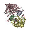
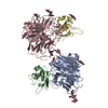
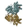
- Structure visualization
| Structure viewer | Molecule: MolmilJmol/JSmol |
|---|
- Downloads & links
-Download
| PDBx/mmCIF format | 3uhj.cif.gz | 521.4 KB | Display | PDBx/mmCIF format |
|---|---|---|---|---|
| PDB format | pdb3uhj.ent.gz | 427.7 KB | Display | PDB format |
| PDBx/mmJSON format | 3uhj.json.gz | Tree view | PDBx/mmJSON format | |
| Others |  Other downloads Other downloads |
-Validation report
| Arichive directory | https://data.pdbj.org/pub/pdb/validation_reports/uh/3uhjftp://data.pdbj.org/pub/pdb/validation_reports/uh/3uhj | HTTPS FTP |
|---|
-Related structure data
| Related structure data | 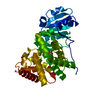 1kq3S S: Starting model for refinement |
|---|---|
| Similar structure data | |
| Other databases |


-Links
 PDBj
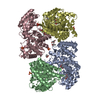
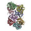
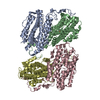
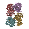
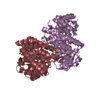
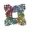
PDBj- Assembly


-Components
| #1: Protein | Mass: 41567.523 Da / Num. of mol.: 8 Source method: isolated from a genetically manipulated source Source: (gene. exp.) Sinorhizobium meliloti (bacteria) / Strain: 1021 / Gene: gldA, R02550, SMc02038 / Plasmid: pET / Production host: #2: Chemical | ChemComp-GOL /   Mass: 92.094 Da / Num. of mol.: 6 / Source method: obtained synthetically / Formula: C3H8O3 Mass: 92.094 Da / Num. of mol.: 6 / Source method: obtained synthetically / Formula: C3H8O3#3: Chemical | ChemComp-ZN /   Mass: 65.409 Da / Num. of mol.: 8 / Source method: obtained synthetically / Formula: Zn Mass: 65.409 Da / Num. of mol.: 8 / Source method: obtained synthetically / Formula: Zn#4: Chemical |   Mass: 78.960 Da / Num. of mol.: 2 / Source method: obtained synthetically / Formula: Se Mass: 78.960 Da / Num. of mol.: 2 / Source method: obtained synthetically / Formula: Se#5: Water | ChemComp-HOH / |  Mass: 18.015 Da / Num. of mol.: 537 / Source method: isolated from a natural source / Formula: H2O Mass: 18.015 Da / Num. of mol.: 537 / Source method: isolated from a natural source / Formula: H2OHas protein modification | Y | |
|---|
-Experimental details
-Experiment
| Experiment | Method: X-RAY DIFFRACTION / Number of used crystals: 1 |
|---|
- Sample preparation
| Crystal | Density Matthews: 2.29 Å3/Da / Density % sol: 46.2 % |
|---|---|
| Crystal grow | Temperature: 293 K / Method: vapor diffusion, sitting drop / pH: 5.5 Details: 0.2M NACL, 0.1M Bis-tris, 25% PEG 3350, 0.2M NDSB 221, pH 5.5, VAPOR DIFFUSION, SITTING DROP, temperature 293K |
-Data collection
| Diffraction | Mean temperature: 100 K |
|---|---|
| Diffraction source | Source: SYNCHROTRON / Site: NSLS  / Beamline: X29A / Wavelength: 0.9792 Å / Beamline: X29A / Wavelength: 0.9792 Å |
| Detector | Type: ADSC QUANTUM 315 / Detector: CCD / Date: Oct 31, 2011 / Details: mirrors |
| Radiation | Monochromator: SI-III / Protocol: SINGLE WAVELENGTH / Monochromatic (M) / Laue (L): M / Scattering type: x-ray |
| Radiation wavelength | Wavelength: 0.9792 Å / Relative weight: 1 |
| Reflection | Resolution: 2.34→50 Å / Num. all: 121795 / Num. obs: 121795 / % possible obs: 98.3 % / Observed criterion σ(I): 0 / Redundancy: 4 % / Rmerge(I) obs: 0.07 / Net I/σ(I): 8.2 |
| Reflection shell | Resolution: 2.34→2.42 Å / Redundancy: 3.6 % / Rmerge(I) obs: 0.5 / Mean I/σ(I) obs: 2.5 / Num. unique all: 11833 / % possible all: 95.8 |
- Processing
| Software |
| |||||||||||||||||||||||||||||||||||||||||||||||||||||||||||||||||
|---|---|---|---|---|---|---|---|---|---|---|---|---|---|---|---|---|---|---|---|---|---|---|---|---|---|---|---|---|---|---|---|---|---|---|---|---|---|---|---|---|---|---|---|---|---|---|---|---|---|---|---|---|---|---|---|---|---|---|---|---|---|---|---|---|---|---|
| Refinement | Method to determine structure: MOLECULAR REPLACEMENT Starting model: 1KQ3 Resolution: 2.34→45.98 Å / Cor.coef. Fo:Fc: 0.947 / Cor.coef. Fo:Fc free: 0.907 / SU B: 8.435 / SU ML: 0.202 / Cross valid method: THROUGHOUT / σ(I): 0 / ESU R: 0.395 / ESU R Free: 0.271 / Stereochemistry target values: MAXIMUM LIKELIHOOD / Details: HYDROGENS HAVE BEEN ADDED IN THE RIDING POSITIONS
| |||||||||||||||||||||||||||||||||||||||||||||||||||||||||||||||||
| Solvent computation | Ion probe radii: 0.8 Å / Shrinkage radii: 0.8 Å / VDW probe radii: 1.4 Å / Solvent model: MASK | |||||||||||||||||||||||||||||||||||||||||||||||||||||||||||||||||
| Displacement parameters | Biso mean: 37.639 Å2
| |||||||||||||||||||||||||||||||||||||||||||||||||||||||||||||||||
| Refinement step | Cycle: LAST / Resolution: 2.34→45.98 Å
| |||||||||||||||||||||||||||||||||||||||||||||||||||||||||||||||||
| Refine LS restraints |
| |||||||||||||||||||||||||||||||||||||||||||||||||||||||||||||||||
| LS refinement shell | Resolution: 2.341→2.402 Å / Total num. of bins used: 20
|
