RNA polymerase II CTD heptapeptide repeat S5 kinase activity / ventricular system development / snRNA transcription by RNA polymerase II / transcription factor TFIIK complex / CAK-ERCC2 complex / transcription factor TFIIH core complex / transcription factor TFIIH holo complex / cyclin-dependent protein serine/threonine kinase activator activity / adult heart development / [RNA-polymerase]-subunit kinase ...RNA polymerase II CTD heptapeptide repeat S5 kinase activity / ventricular system development / snRNA transcription by RNA polymerase II / transcription factor TFIIK complex / CAK-ERCC2 complex / transcription factor TFIIH core complex / transcription factor TFIIH holo complex / cyclin-dependent protein serine/threonine kinase activator activity / adult heart development / [RNA-polymerase]-subunit kinase / RNA Polymerase I Transcription Termination / cyclin-dependent protein serine/threonine kinase regulator activity / RNA Pol II CTD phosphorylation and interaction with CE during HIV infection / RNA Pol II CTD phosphorylation and interaction with CE / Formation of the Early Elongation Complex / Formation of the HIV-1 Early Elongation Complex / mRNA Capping / HIV Transcription Initiation / RNA Polymerase II HIV Promoter Escape / Transcription of the HIV genome / RNA Polymerase II Promoter Escape / RNA Polymerase II Transcription Pre-Initiation And Promoter Opening / RNA Polymerase II Transcription Initiation / RNA Polymerase II Transcription Initiation And Promoter Clearance / RNA Polymerase I Transcription Initiation / RNA polymerase II transcribes snRNA genes / regulation of G1/S transition of mitotic cell cycle / cyclin-dependent kinase / cyclin-dependent protein serine/threonine kinase activity / ATP-dependent activity, acting on DNA / Tat-mediated elongation of the HIV-1 transcript / Formation of HIV-1 elongation complex containing HIV-1 Tat / Cyclin E associated events during G1/S transition / Formation of HIV elongation complex in the absence of HIV Tat / Cyclin A:Cdk2-associated events at S phase entry / cyclin-dependent protein kinase holoenzyme complex / Cyclin A/B1/B2 associated events during G2/M transition / RNA Polymerase II Transcription Elongation / Formation of RNA Pol II elongation complex / RNA polymerase II CTD heptapeptide repeat kinase activity / RNA Polymerase II Pre-transcription Events / positive regulation of smooth muscle cell proliferation / male germ cell nucleus / TP53 Regulates Transcription of DNA Repair Genes / nucleotide-excision repair / RNA Polymerase I Promoter Escape / transcription initiation at RNA polymerase II promoter / G1/S transition of mitotic cell cycle / NoRC negatively regulates rRNA expression / response to calcium ion / fibrillar center / Transcription-Coupled Nucleotide Excision Repair (TC-NER) / Formation of TC-NER Pre-Incision Complex / Formation of Incision Complex in GG-NER / kinase activity / Cyclin D associated events in G1 / Dual incision in TC-NER / Gap-filling DNA repair synthesis and ligation in TC-NER / RUNX1 regulates transcription of genes involved in differentiation of HSCs / transcription by RNA polymerase II / protein kinase activity / regulation of cell cycle / protein stabilization / protein serine kinase activity / cell division / DNA repair / protein serine/threonine kinase activity / regulation of transcription by RNA polymerase II / negative regulation of apoptotic process / perinuclear region of cytoplasm / positive regulation of transcription by RNA polymerase II / zinc ion binding / nucleoplasm / ATP binding / nucleus / plasma membrane / cytoplasm / cytosol Similarity search - Function
: / MAT1 C-terminal CAK anchor / CyclinH/Ccl1 / Cyclin-dependent kinase 7 / Cdk-activating kinase assembly factor MAT1/Tfb3 / Cdk-activating kinase assembly factor MAT1, centre / CDK-activating kinase assembly factor MAT1 / Zinc finger, C3HC4 type (RING finger) / Cyclin, C-terminal domain 2 / Cyclin C-terminal domain ...: / MAT1 C-terminal CAK anchor / CyclinH/Ccl1 / Cyclin-dependent kinase 7 / Cdk-activating kinase assembly factor MAT1/Tfb3 / Cdk-activating kinase assembly factor MAT1, centre / CDK-activating kinase assembly factor MAT1 / Zinc finger, C3HC4 type (RING finger) / Cyclin, C-terminal domain 2 / Cyclin C-terminal domain / Cyclin/Cyclin-like subunit Ssn8 / Cyclin, N-terminal / Cyclin, N-terminal domain / Ubiquitin interacting motif / Ubiquitin-interacting motif (UIM) domain profile. / Cyclin-like / domain present in cyclins, TFIIB and Retinoblastoma / Cyclin-like superfamily / : / Zinc finger, RING-type, conserved site / Zinc finger RING-type signature. / Ring finger / Zinc finger RING-type profile. / Zinc finger, RING-type / Zinc finger, RING/FYVE/PHD-type / Serine/threonine-protein kinase, active site / Serine/Threonine protein kinases active-site signature. / Protein kinase domain / Serine/Threonine protein kinases, catalytic domain / Protein kinase, ATP binding site / Protein kinases ATP-binding region signature. / Protein kinase domain profile. / Protein kinase domain / Protein kinase-like domain superfamily Similarity search - Domain/homology
National Institutes of Health/National Institute of General Medical Sciences (NIH/NIGMS)
R01-GM63072
United States
National Institutes of Health/National Institute of General Medical Sciences (NIH/NIGMS)
P01-GM063210
United States
National Institutes of Health/National Institute of General Medical Sciences (NIH/NIGMS)
R35-GM127018
United States
Howard Hughes Medical Institute (HHMI)
United States
Citation
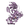
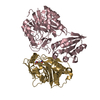
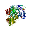
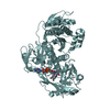
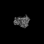
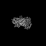
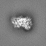
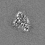
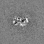
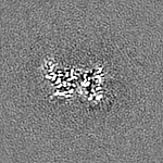
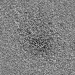
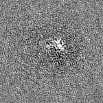
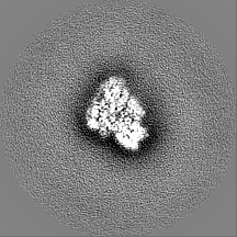
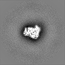
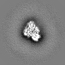
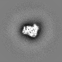
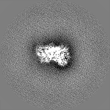
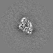
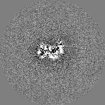
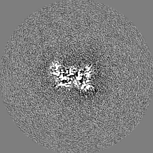
Journal: Proc Natl Acad Sci U S A / Year: 2020 Title: The cryoelectron microscopy structure of the human CDK-activating kinase. Authors: Basil J Greber / Juan M Perez-Bertoldi / Kif Lim / Anthony T Iavarone / Daniel B Toso / Eva Nogales / Abstract: The human CDK-activating kinase (CAK), a complex composed of cyclin-dependent kinase (CDK) 7, cyclin H, and MAT1, is a critical regulator of transcription initiation and the cell cycle. It acts by ...The human CDK-activating kinase (CAK), a complex composed of cyclin-dependent kinase (CDK) 7, cyclin H, and MAT1, is a critical regulator of transcription initiation and the cell cycle. It acts by phosphorylating the C-terminal heptapeptide repeat domain of the RNA polymerase II (Pol II) subunit RPB1, which is an important regulatory event in transcription initiation by Pol II, and it phosphorylates the regulatory T-loop of CDKs that control cell cycle progression. Here, we have determined the three-dimensional (3D) structure of the catalytic module of human CAK, revealing the structural basis of its assembly and providing insight into CDK7 activation in this context. The unique third component of the complex, MAT1, substantially extends the interaction interface between CDK7 and cyclin H, explaining its role as a CAK assembly factor, and it forms interactions with the CDK7 T-loop, which may contribute to enhancing CAK activity. We have also determined the structure of the CAK in complex with the covalently bound inhibitor THZ1 in order to provide insight into the binding of inhibitors at the CDK7 active site and to aid in the rational design of therapeutic compounds.
History
Deposition
Jun 7, 2020
-
Header (metadata) release
Sep 9, 2020
-
Map release
Sep 9, 2020
-
Update
Nov 6, 2024
-
Current status
Nov 6, 2024
Processing site: RCSB / Status: Released
-
Structure visualization
Movie
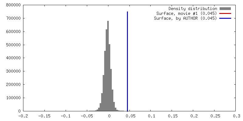
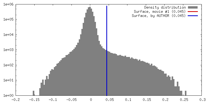
Surface view with section colored by density value
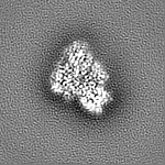
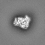
EMPIAR-10431 (Title: Single-particle cryo-EM of the human CDK-activating kinase in complex with ATP-gamma-S Data size: 3.5 TB Data #1: Unaligned movies of human CAK, dataset 1 [micrographs - multiframe] Data #2: Unaligned movies of human CAK, dataset 2 [micrographs - multiframe])
Model: UltrAuFoil R1.2/1.3 / Material: GOLD / Support film - Material: GOLD / Support film - topology: HOLEY / Pretreatment - Type: PLASMA CLEANING / Pretreatment - Time: 30 sec. / Pretreatment - Atmosphere: AIR
Vitrification
Cryogen name: ETHANE / Chamber humidity: 100 % / Chamber temperature: 278 K / Instrument: FEI VITROBOT MARK IV
-
Electron microscopy
Microscope
FEI TALOS ARCTICA
Image recording
#0 - Image recording ID: 1 / #0 - Film or detector model: GATAN K3 (6k x 4k) / #0 - Number grids imaged: 1 / #0 - Number real images: 3296 / #0 - Average exposure time: 2.0 sec. / #0 - Average electron dose: 69.0 e/Å2 / #0 - Details: 69 frames per movie / #1 - Image recording ID: 2 / #1 - Film or detector model: GATAN K3 (6k x 4k) / #1 - Number grids imaged: 1 / #1 - Number real images: 4286 / #1 - Average exposure time: 2.0 sec. / #1 - Average electron dose: 69.0 e/Å2 / #1 - Details: 69 frames per movie
Electron beam
Acceleration voltage: 200 kV / Electron source: FIELD EMISSION GUN
In the structure databanks used in Yorodumi, some data are registered as the other names, "COVID-19 virus" and "2019-nCoV". Here are the details of the virus and the list of structure data.
Jan 31, 2019. EMDB accession codes are about to change! (news from PDBe EMDB page)
EMDB accession codes are about to change! (news from PDBe EMDB page)
The allocation of 4 digits for EMDB accession codes will soon come to an end. Whilst these codes will remain in use, new EMDB accession codes will include an additional digit and will expand incrementally as the available range of codes is exhausted. The current 4-digit format prefixed with “EMD-” (i.e. EMD-XXXX) will advance to a 5-digit format (i.e. EMD-XXXXX), and so on. It is currently estimated that the 4-digit codes will be depleted around Spring 2019, at which point the 5-digit format will come into force.
The EM Navigator/Yorodumi systems omit the EMD- prefix.
Related info.:Q: What is EMD? / ID/Accession-code notation in Yorodumi/EM Navigator
Yorodumi is a browser for structure data from EMDB, PDB, SASBDB, etc.
This page is also the successor to EM Navigator detail page, and also detail information page/front-end page for Omokage search.
The word "yorodu" (or yorozu) is an old Japanese word meaning "ten thousand". "mi" (miru) is to see.
Related info.:EMDB / PDB / SASBDB / Comparison of 3 databanks / Yorodumi Search / Aug 31, 2016. New EM Navigator & Yorodumi / Yorodumi Papers / Jmol/JSmol / Function and homology information / Changes in new EM Navigator and Yorodumi
 Movie
Movie Controller


 Open data
 Basic information
 Map data
 Sample
 Keywords
 Function and homology information
 Homo sapiens (human)
 Authors
 United States, 4 items
  Citation
 Structure visualization
 Downloads & links
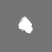
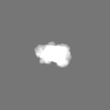
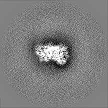
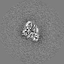
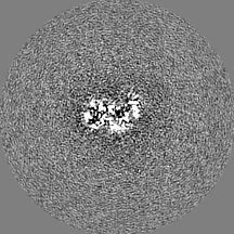
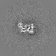
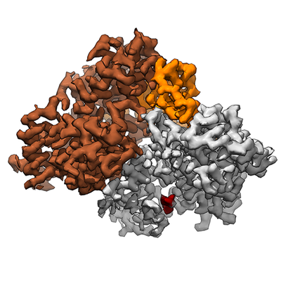 emd_22123.png
 http://ftp.pdbj.org/pub/emdb/structures/EMD-22123


 Z (Sec.)
 Y (Row.)
 X (Col.)


 Sample components
 Trichoplusia ni (cabbage looper)
 Processing
 Electron microscopy
 FIELD EMISSION GUN


