 Movie
Movie Controller


+ Open data

- Basic information
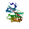
| Entry | Database: PDB / ID: 1ua2 | ||||||
|---|---|---|---|---|---|---|---|
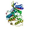
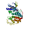
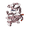
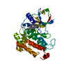
| Title | Crystal Structure of Human CDK7 | ||||||
 Components Components | Cell division protein kinase 7 | ||||||
 Keywords Keywords | CELL CYCLE / TRANSFERASE / PHOSPHORYLATION / PROTEIN-PROTEIN INTERACTION / PROTEIN KINASE | ||||||
| Function / homology |  Function and homology information Function and homology informationRNA polymerase II CTD heptapeptide repeat S5 kinase activity / snRNA transcription by RNA polymerase II / transcription factor TFIIK complex / CAK-ERCC2 complex / transcription factor TFIIH core complex / transcription factor TFIIH holo complex / [RNA-polymerase]-subunit kinase / RNA Polymerase I Transcription Termination / RNA Pol II CTD phosphorylation and interaction with CE during HIV infection / RNA Pol II CTD phosphorylation and interaction with CE ...RNA polymerase II CTD heptapeptide repeat S5 kinase activity / snRNA transcription by RNA polymerase II / transcription factor TFIIK complex / CAK-ERCC2 complex / transcription factor TFIIH core complex / transcription factor TFIIH holo complex / [RNA-polymerase]-subunit kinase / RNA Polymerase I Transcription Termination / RNA Pol II CTD phosphorylation and interaction with CE during HIV infection / RNA Pol II CTD phosphorylation and interaction with CE / Formation of the Early Elongation Complex / Formation of the HIV-1 Early Elongation Complex / mRNA Capping / HIV Transcription Initiation / RNA Polymerase II HIV Promoter Escape / Transcription of the HIV genome / RNA Polymerase II Promoter Escape / RNA Polymerase II Transcription Pre-Initiation And Promoter Opening / RNA Polymerase II Transcription Initiation / RNA Polymerase II Transcription Initiation And Promoter Clearance / RNA Polymerase I Transcription Initiation / RNA polymerase II transcribes snRNA genes / cyclin-dependent kinase / cyclin-dependent protein serine/threonine kinase activity / ATP-dependent activity, acting on DNA / Tat-mediated elongation of the HIV-1 transcript / Formation of HIV-1 elongation complex containing HIV-1 Tat / Cyclin E associated events during G1/S transition / Cyclin A:Cdk2-associated events at S phase entry / Formation of HIV elongation complex in the absence of HIV Tat / Cyclin A/B1/B2 associated events during G2/M transition / cyclin-dependent protein kinase holoenzyme complex / RNA Polymerase II Transcription Elongation / regulation of G1/S transition of mitotic cell cycle / Formation of RNA Pol II elongation complex / RNA polymerase II CTD heptapeptide repeat kinase activity / RNA Polymerase II Pre-transcription Events / male germ cell nucleus / TP53 Regulates Transcription of DNA Repair Genes / RNA Polymerase I Promoter Escape / transcription initiation at RNA polymerase II promoter / NoRC negatively regulates rRNA expression / fibrillar center / Transcription-Coupled Nucleotide Excision Repair (TC-NER) / Formation of TC-NER Pre-Incision Complex / Formation of Incision Complex in GG-NER / Cyclin D associated events in G1 / Dual incision in TC-NER / Gap-filling DNA repair synthesis and ligation in TC-NER / RUNX1 regulates transcription of genes involved in differentiation of HSCs / transcription by RNA polymerase II / protein kinase activity / regulation of cell cycle / protein stabilization / protein serine kinase activity / cell division / DNA repair / protein serine/threonine kinase activity / perinuclear region of cytoplasm / positive regulation of transcription by RNA polymerase II / nucleoplasm / ATP binding / nucleus / plasma membrane / cytosol / cytoplasm Similarity search - Function | ||||||
| Biological species |  Homo sapiens (human) Homo sapiens (human) | ||||||
| Method |  X-RAY DIFFRACTION / SYNCHROTRON / MOLECULAR REPLACEMENT / Resolution: 3.02 Å X-RAY DIFFRACTION / SYNCHROTRON / MOLECULAR REPLACEMENT / Resolution: 3.02 Å | ||||||
 Authors Authors | Lolli, G. / Lowe, E.D. / Brown, N.R. / Johnson, L.N. | ||||||
 Citation Citation | Journal: Structure / Year: 2004 Title: The Crystal Structure of Human CDK7 and Its Protein Recognition Properties Authors: Lolli, G. / Lowe, E.D. / Brown, N.R. / Johnson, L.N. | ||||||
| History |
|
- Structure visualization
| Structure viewer | Molecule: MolmilJmol/JSmol |
|---|
- Downloads & links
-Download
| PDBx/mmCIF format | 1ua2.cif.gz | 239.7 KB | Display | PDBx/mmCIF format |
|---|---|---|---|---|
| PDB format | pdb1ua2.ent.gz | 193.3 KB | Display | PDB format |
| PDBx/mmJSON format | 1ua2.json.gz | Tree view | PDBx/mmJSON format | |
| Others |  Other downloads Other downloads |
-Validation report
| Arichive directory | https://data.pdbj.org/pub/pdb/validation_reports/ua/1ua2ftp://data.pdbj.org/pub/pdb/validation_reports/ua/1ua2 | HTTPS FTP |
|---|
-Related structure data
| Similar structure data |
|---|


-Links
 PDBj
PDBj


- Assembly
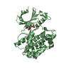
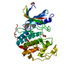
| Deposited unit | 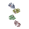
| ||||||||||||||||||||||||||||||
|---|---|---|---|---|---|---|---|---|---|---|---|---|---|---|---|---|---|---|---|---|---|---|---|---|---|---|---|---|---|---|---|
| 1 | 
| ||||||||||||||||||||||||||||||
| 2 | 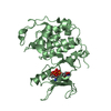
| ||||||||||||||||||||||||||||||
| 3 | 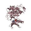
| ||||||||||||||||||||||||||||||
| 4 | 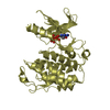
| ||||||||||||||||||||||||||||||
| Unit cell |
| ||||||||||||||||||||||||||||||
| Noncrystallographic symmetry (NCS) | NCS domain:
NCS domain segments: Component-ID: 1 / Ens-ID: 1 / Beg auth comp-ID: GLU / Beg label comp-ID: GLU / End auth comp-ID: ASN / End label comp-ID: ASN / Refine code: 6 / Auth seq-ID: 13 - 311 / Label seq-ID: 13 - 311
| ||||||||||||||||||||||||||||||
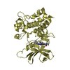
| Details | The biological unit is a monomer . The presence of four copies in the assymetric unit has no biological signifigance |
-Components
| #1: Protein | Mass: 39170.324 Da / Num. of mol.: 4 Source method: isolated from a genetically manipulated source Source: (gene. exp.) Homo sapiens (human) / Description: Sf21 cells in culture / Gene: CDK7 / Plasmid: pVL1392 / Cell line (production host): Sf21 / Production host:   Spodoptera frugiperda (fall armyworm) / References: UniProt: P50613, EC: 2.7.1.37 Spodoptera frugiperda (fall armyworm) / References: UniProt: P50613, EC: 2.7.1.37#2: Chemical | ChemComp-ATP /   Mass: 507.181 Da / Num. of mol.: 4 / Source method: obtained synthetically / Formula: C10H16N5O13P3 / Comment: ATP, energy-carrying molecule*YM Mass: 507.181 Da / Num. of mol.: 4 / Source method: obtained synthetically / Formula: C10H16N5O13P3 / Comment: ATP, energy-carrying molecule*YM#3: Water | ChemComp-HOH / |  Mass: 18.015 Da / Num. of mol.: 48 / Source method: isolated from a natural source / Formula: H2O Mass: 18.015 Da / Num. of mol.: 48 / Source method: isolated from a natural source / Formula: H2OHas protein modification | Y | |
|---|
-Experimental details
-Experiment
| Experiment | Method: X-RAY DIFFRACTION / Number of used crystals: 2 |
|---|
- Sample preparation
| Crystal | Density Matthews: 3.63 Å3/Da / Density % sol: 65.8 % |
|---|---|
| Crystal grow | Temperature: 277 K / Method: vapor diffusion, sitting drop / pH: 6.4 Details: Na Citrate, Na Acetate, PEG 4000, glycerol, NDSB201, ATP, pH 6.4, VAPOR DIFFUSION, SITTING DROP, temperature 4.0K |
-Data collection
| Diffraction |
| |||||||||||||||
|---|---|---|---|---|---|---|---|---|---|---|---|---|---|---|---|---|
| Diffraction source |
| |||||||||||||||
| Detector |
| |||||||||||||||
| Radiation | Protocol: SINGLE WAVELENGTH / Monochromatic (M) / Laue (L): M / Scattering type: x-ray | |||||||||||||||
| Radiation wavelength |
| |||||||||||||||
| Reflection | Resolution: 2.96→49.45 Å / Num. all: 38122 / Num. obs: 35416 / % possible obs: 92.9 % / Observed criterion σ(I): 1 / Redundancy: 3.8 % / Biso Wilson estimate: 73.1 Å2 / Rmerge(I) obs: 0.129 / Rsym value: 0.129 / Net I/σ(I): 9.5 | |||||||||||||||
| Reflection shell | Resolution: 3.02→3.18 Å / Redundancy: 1.7 % / Rmerge(I) obs: 0.466 / Mean I/σ(I) obs: 1.6 / Num. unique all: 3851 / Rsym value: 0.466 / % possible all: 70.1 |

- Processing
| Software |
| |||||||||||||||||||||||||||||||||||||||||||||||||||||||||||||||||||||||||||||||||||||||||||||||||||||||||||||||||||||||||||||
|---|---|---|---|---|---|---|---|---|---|---|---|---|---|---|---|---|---|---|---|---|---|---|---|---|---|---|---|---|---|---|---|---|---|---|---|---|---|---|---|---|---|---|---|---|---|---|---|---|---|---|---|---|---|---|---|---|---|---|---|---|---|---|---|---|---|---|---|---|---|---|---|---|---|---|---|---|---|---|---|---|---|---|---|---|---|---|---|---|---|---|---|---|---|---|---|---|---|---|---|---|---|---|---|---|---|---|---|---|---|---|---|---|---|---|---|---|---|---|---|---|---|---|---|---|---|---|
| Refinement | Method to determine structure: MOLECULAR REPLACEMENT Starting model: Human CDK2 Resolution: 3.02→49.45 Å / Cor.coef. Fo:Fc: 0.911 / Cor.coef. Fo:Fc free: 0.844 / SU B: 42.022 / SU ML: 0.364 / TLS residual ADP flag: LIKELY RESIDUAL / Cross valid method: THROUGHOUT / σ(F): 0 / ESU R Free: 0.473 / Stereochemistry target values: MAXIMUM LIKELIHOOD / Details: HYDROGENS HAVE BEEN ADDED IN THE RIDING POSITIONS
| |||||||||||||||||||||||||||||||||||||||||||||||||||||||||||||||||||||||||||||||||||||||||||||||||||||||||||||||||||||||||||||
| Solvent computation | Ion probe radii: 0.8 Å / Shrinkage radii: 0.8 Å / VDW probe radii: 1.2 Å / Solvent model: BABINET MODEL WITH MASK | |||||||||||||||||||||||||||||||||||||||||||||||||||||||||||||||||||||||||||||||||||||||||||||||||||||||||||||||||||||||||||||
| Displacement parameters | Biso mean: 44.013 Å2
| |||||||||||||||||||||||||||||||||||||||||||||||||||||||||||||||||||||||||||||||||||||||||||||||||||||||||||||||||||||||||||||
| Refinement step | Cycle: LAST / Resolution: 3.02→49.45 Å
| |||||||||||||||||||||||||||||||||||||||||||||||||||||||||||||||||||||||||||||||||||||||||||||||||||||||||||||||||||||||||||||
| Refine LS restraints |
| |||||||||||||||||||||||||||||||||||||||||||||||||||||||||||||||||||||||||||||||||||||||||||||||||||||||||||||||||||||||||||||
| Refine LS restraints NCS | Ens-ID: 1 / Number: 2291 / Refine-ID: X-RAY DIFFRACTION
| |||||||||||||||||||||||||||||||||||||||||||||||||||||||||||||||||||||||||||||||||||||||||||||||||||||||||||||||||||||||||||||
| LS refinement shell | Resolution: 3.02→3.078 Å / Total num. of bins used: 20
| |||||||||||||||||||||||||||||||||||||||||||||||||||||||||||||||||||||||||||||||||||||||||||||||||||||||||||||||||||||||||||||
| Refinement TLS params. | Method: refined / Refine-ID: X-RAY DIFFRACTION
| |||||||||||||||||||||||||||||||||||||||||||||||||||||||||||||||||||||||||||||||||||||||||||||||||||||||||||||||||||||||||||||
| Refinement TLS group |
|
