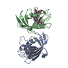
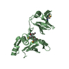
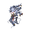
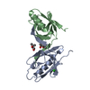
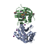
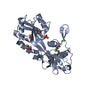
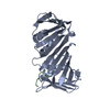
- PDB-3uen: Crystal structure of TopBP1 BRCT4/5 domains -
+
データを開く
IDまたはキーワード:
読み込み中...
-
基本情報
登録情報
データベース: PDB / ID: 3uen
タイトル
Crystal structure of TopBP1 BRCT4/5 domains
要素
DNA topoisomerase 2-binding protein 1
キーワード
PEPTIDE BINDING PROTEIN / BRCT domain / phospho-peptide binding
機能・相同性
機能・相同性情報
broken chromosome clustering / BRCA1-B complex / phosphorylation-dependent protein binding / homologous recombination / DNA replication checkpoint signaling / double-strand break repair via classical nonhomologous end joining / double-strand break repair via alternative nonhomologous end joining / mitotic DNA replication checkpoint signaling / protein localization to site of double-strand break / chromatin-protein adaptor activity ...broken chromosome clustering / BRCA1-B complex / phosphorylation-dependent protein binding / homologous recombination / DNA replication checkpoint signaling / double-strand break repair via classical nonhomologous end joining / double-strand break repair via alternative nonhomologous end joining / mitotic DNA replication checkpoint signaling / protein localization to site of double-strand break / chromatin-protein adaptor activity / HDR through Single Strand Annealing (SSA) / DNA metabolic process / response to ionizing radiation / mitotic G2 DNA damage checkpoint signaling / Impaired BRCA2 binding to RAD51 / Presynaptic phase of homologous DNA pairing and strand exchange / chromosome organization / DNA replication initiation / site of DNA damage / DNA damage checkpoint signaling / condensed nuclear chromosome / male germ cell nucleus / protein serine/threonine kinase activator activity / PML body / G2/M DNA damage checkpoint / double-strand break repair via homologous recombination / spindle pole / chromosome / actin cytoskeleton / site of double-strand break / Processing of DNA double-strand break ends / Regulation of TP53 Activity through Phosphorylation / nuclear body / DNA repair / DNA damage response / centrosome / DNA binding / nucleoplasm / identical protein binding / nucleus / plasma membrane 類似検索 - 分子機能
 ムービー
ムービー コントローラー


 データを開く
 基本情報
基本情報 要素
要素 キーワード
キーワード 機能・相同性情報
 Homo sapiens (ヒト)
 X線回折 /
  データ登録者
 引用
引用 構造の表示
構造の表示 ダウンロードとリンク
 その他のダウンロード


 PDBj
PDBj


 集合体
集合体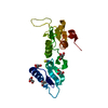


 分子量: 58.082 Da / 分子数: 1 / 由来タイプ: 合成 / 式: CNS
 分子量: 92.094 Da / 分子数: 5 / 由来タイプ: 合成 / 式: C3H8O3
 分子量: 18.015 Da / 分子数: 238 / 由来タイプ: 天然 / 式: H2O
 試料調製
試料調製 / ビームライン: 8.3.1 / 波長: 1.11588 Å
 解析
解析