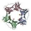
 Movie
Movie Controller


+ Open data

- Basic information
| Entry | Database: PDB / ID: 6t7y | ||||||
|---|---|---|---|---|---|---|---|
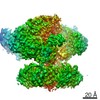
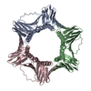
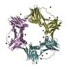
| Title | Structure of PCNA bound to cPIP motif of DP2 from P. abyssi | ||||||
 Components Components |
| ||||||
 Keywords Keywords | REPLICATION / PCNA / DP2 / PolD / PIP-box | ||||||
| Function / homology |  Function and homology information Function and homology informationexodeoxyribonuclease I / intein-mediated protein splicing / single-stranded DNA 3'-5' DNA exonuclease activity / DNA catabolic process / leading strand elongation / DNA polymerase processivity factor activity / regulation of DNA replication / DNA-templated DNA replication / DNA-directed DNA polymerase / DNA-directed DNA polymerase activity ...exodeoxyribonuclease I / intein-mediated protein splicing / single-stranded DNA 3'-5' DNA exonuclease activity / DNA catabolic process / leading strand elongation / DNA polymerase processivity factor activity / regulation of DNA replication / DNA-templated DNA replication / DNA-directed DNA polymerase / DNA-directed DNA polymerase activity / DNA binding / identical protein binding Similarity search - Function | ||||||
| Biological species |   Pyrococcus abyssi (archaea) Pyrococcus abyssi (archaea) | ||||||
| Method |  X-RAY DIFFRACTION / SYNCHROTRON / MOLECULAR REPLACEMENT / molecular replacement / Resolution: 2.7 Å X-RAY DIFFRACTION / SYNCHROTRON / MOLECULAR REPLACEMENT / molecular replacement / Resolution: 2.7 Å | ||||||
 Authors Authors | Madru, C. / Raia, P. / Hugonneau Beaufet, I. / Delarue, M. / Carroni, M. / Sauguet, L. | ||||||
| Funding support |  France, 1items France, 1items
| ||||||
 Citation Citation | Journal: Nat Commun / Year: 2020 Title: Structural basis for the increased processivity of D-family DNA polymerases in complex with PCNA. Authors: Clément Madru / Ghislaine Henneke / Pierre Raia / Inès Hugonneau-Beaufet / Gérard Pehau-Arnaudet / Patrick England / Erik Lindahl / Marc Delarue / Marta Carroni / Ludovic Sauguet /  Abstract: Replicative DNA polymerases (DNAPs) have evolved the ability to copy the genome with high processivity and fidelity. In Eukarya and Archaea, the processivity of replicative DNAPs is greatly enhanced ...Replicative DNA polymerases (DNAPs) have evolved the ability to copy the genome with high processivity and fidelity. In Eukarya and Archaea, the processivity of replicative DNAPs is greatly enhanced by its binding to the proliferative cell nuclear antigen (PCNA) that encircles the DNA. We determined the cryo-EM structure of the DNA-bound PolD-PCNA complex from Pyrococcus abyssi at 3.77 Å. Using an integrative structural biology approach - combining cryo-EM, X-ray crystallography, protein-protein interaction measurements, and activity assays - we describe the molecular basis for the interaction and cooperativity between a replicative DNAP and PCNA. PolD recruits PCNA via a complex mechanism, which requires two different PIP-boxes. We infer that the second PIP-box, which is shared with the eukaryotic Polα replicative DNAP, plays a dual role in binding either PCNA or primase, and could be a master switch between an initiation and a processive phase during replication. | ||||||
| History |
|
- Structure visualization
| Structure viewer | Molecule: MolmilJmol/JSmol |
|---|
- Downloads & links
-Download
| PDBx/mmCIF format | 6t7y.cif.gz | 113.5 KB | Display | PDBx/mmCIF format |
|---|---|---|---|---|
| PDB format | pdb6t7y.ent.gz | 88.2 KB | Display | PDB format |
| PDBx/mmJSON format | 6t7y.json.gz | Tree view | PDBx/mmJSON format | |
| Others |  Other downloads Other downloads |
-Validation report
| Arichive directory | https://data.pdbj.org/pub/pdb/validation_reports/t7/6t7yftp://data.pdbj.org/pub/pdb/validation_reports/t7/6t7y | HTTPS FTP |
|---|
-Related structure data
| Related structure data |  6t7xC 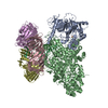 6t8hC  5aujS S: Starting model for refinement C: citing same article ( |
|---|---|
| Similar structure data |


-Links
 PDBj
PDBj


- Assembly
| Deposited unit | 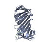
| ||||||||
|---|---|---|---|---|---|---|---|---|---|
| 1 | 
| ||||||||
| Unit cell |
|
-Components
| #1: Protein | Mass: 29471.764 Da / Num. of mol.: 1 Source method: isolated from a genetically manipulated source Source: (gene. exp.) Pyrococcus abyssi (strain GE5 / Orsay) (archaea)Strain: GE5 / Orsay / Gene: pcn, PYRAB13790, PAB1465 / Production host:  |
|---|---|
| #2: Protein/peptide | Mass: 1485.746 Da / Num. of mol.: 1 Source method: isolated from a genetically manipulated source Source: (gene. exp.) Pyrococcus abyssi (archaea) / Production host: synthetic construct (others)References: UniProt: Q9V2F4*PLUS, DNA-directed DNA polymerase |
| #3: Water | ChemComp-HOH /  Mass: 18.015 Da / Num. of mol.: 14 / Source method: isolated from a natural source / Formula: H2O Mass: 18.015 Da / Num. of mol.: 14 / Source method: isolated from a natural source / Formula: H2O |
-Experimental details
-Experiment
| Experiment | Method: X-RAY DIFFRACTION / Number of used crystals: 1 |
|---|
- Sample preparation
| Crystal | Density Matthews: 2.37 Å3/Da / Density % sol: 48.13 % |
|---|---|
| Crystal grow | Temperature: 293 K / Method: vapor diffusion, sitting drop / pH: 7.1 / Details: 30% PEG 400 0.2 M MgCl2 0.1M BIS-TRIS pH7.1 |
-Data collection
| Diffraction | Mean temperature: 100 K / Serial crystal experiment: N |
|---|---|
| Diffraction source | Source: SYNCHROTRON / Site: ESRF / Beamline: ID29 / Wavelength: 0.9793 Å |
| Detector | Type: DECTRIS PILATUS 6M / Detector: PIXEL / Date: Nov 11, 2018 |
| Radiation | Protocol: SINGLE WAVELENGTH / Monochromatic (M) / Laue (L): M / Scattering type: x-ray |
| Radiation wavelength | Wavelength: 0.9793 Å / Relative weight: 1 |
| Reflection | Resolution: 2.7→70.37 Å / Num. obs: 6983 / % possible obs: 89.5 % / Redundancy: 5.2 % / CC1/2: 0.999 / Net I/σ(I): 15.8 |
| Reflection shell | Resolution: 2.701→2.863 Å / Num. unique obs: 436 / CC1/2: 0.21 |
-Phasing
| Phasing | Method: molecular replacement |
|---|
- Processing
| Software |
| |||||||||||||||||||||||||||||||||||||||||||||||||||||||||||||||||||||||||||
|---|---|---|---|---|---|---|---|---|---|---|---|---|---|---|---|---|---|---|---|---|---|---|---|---|---|---|---|---|---|---|---|---|---|---|---|---|---|---|---|---|---|---|---|---|---|---|---|---|---|---|---|---|---|---|---|---|---|---|---|---|---|---|---|---|---|---|---|---|---|---|---|---|---|---|---|---|
| Refinement | Method to determine structure: MOLECULAR REPLACEMENT Starting model: 5auj Resolution: 2.7→70.37 Å / Cor.coef. Fo:Fc: 0.966 / Cor.coef. Fo:Fc free: 0.937 / SU B: 50.167 / SU ML: 0.427 / Cross valid method: THROUGHOUT / σ(F): 0 / ESU R Free: 0.424 Details: HYDROGENS HAVE BEEN ADDED IN THE RIDING POSITIONS U VALUES : WITH TLS ADDED
| |||||||||||||||||||||||||||||||||||||||||||||||||||||||||||||||||||||||||||
| Solvent computation | Ion probe radii: 0.8 Å / Shrinkage radii: 0.8 Å / VDW probe radii: 1.2 Å | |||||||||||||||||||||||||||||||||||||||||||||||||||||||||||||||||||||||||||
| Displacement parameters | Biso max: 218.27 Å2 / Biso mean: 134.05 Å2 / Biso min: 84.66 Å2
| |||||||||||||||||||||||||||||||||||||||||||||||||||||||||||||||||||||||||||
| Refinement step | Cycle: final / Resolution: 2.7→70.37 Å
| |||||||||||||||||||||||||||||||||||||||||||||||||||||||||||||||||||||||||||
| Refine LS restraints |
| |||||||||||||||||||||||||||||||||||||||||||||||||||||||||||||||||||||||||||
| LS refinement shell | Resolution: 2.701→2.771 Å / Rfactor Rfree error: 0 / Total num. of bins used: 20
| |||||||||||||||||||||||||||||||||||||||||||||||||||||||||||||||||||||||||||
| Refinement TLS params. | Method: refined / Refine-ID: X-RAY DIFFRACTION
| |||||||||||||||||||||||||||||||||||||||||||||||||||||||||||||||||||||||||||
| Refinement TLS group |
|
