 Movie
Movie Controller


+ Open data

- Basic information
| Entry | Database: PDB / ID: 1isq | ||||||
|---|---|---|---|---|---|---|---|
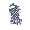
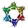
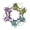
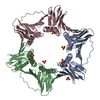
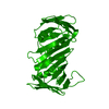
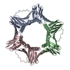
| Title | Pyrococcus furiosus PCNA complexed with RFCL PIP-box peptide | ||||||
 Components Components |
| ||||||
 Keywords Keywords | DNA BINDING PROTEIN / Toroidal trimer | ||||||
| Function / homology |  Function and homology information Function and homology informationDNA clamp loader activity / leading strand elongation / DNA polymerase processivity factor activity / regulation of DNA replication / DNA replication / ATP hydrolysis activity / DNA binding / ATP binding / identical protein binding Similarity search - Function | ||||||
| Biological species |   Pyrococcus furiosus (archaea) Pyrococcus furiosus (archaea) | ||||||
| Method |  X-RAY DIFFRACTION / SYNCHROTRON / MOLECULAR REPLACEMENT / Resolution: 2.3 Å X-RAY DIFFRACTION / SYNCHROTRON / MOLECULAR REPLACEMENT / Resolution: 2.3 Å | ||||||
 Authors Authors | Matsumiya, S. / Ishino, S. / Ishino, Y. / Morikawa, K. | ||||||
 Citation Citation | Journal: Genes Cells / Year: 2002 Title: Physical interaction between proliferating cell nuclear antigen and replication factor C from Pyrococcus furiosus Authors: Matsumiya, S. / Ishino, S. / Ishino, Y. / Morikawa, K. | ||||||
| History |
|
- Structure visualization
| Structure viewer | Molecule: MolmilJmol/JSmol |
|---|
- Downloads & links
-Download
| PDBx/mmCIF format | 1isq.cif.gz | 60.8 KB | Display | PDBx/mmCIF format |
|---|---|---|---|---|
| PDB format | pdb1isq.ent.gz | 44.9 KB | Display | PDB format |
| PDBx/mmJSON format | 1isq.json.gz | Tree view | PDBx/mmJSON format | |
| Others |  Other downloads Other downloads |
-Validation report
| Arichive directory | https://data.pdbj.org/pub/pdb/validation_reports/is/1isqftp://data.pdbj.org/pub/pdb/validation_reports/is/1isq | HTTPS FTP |
|---|
-Related structure data
| Related structure data |  1ge8S S: Starting model for refinement |
|---|---|
| Similar structure data |


-Links
 PDBj
PDBj

- Assembly
| Deposited unit | 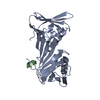
| ||||||||
|---|---|---|---|---|---|---|---|---|---|
| 1 | 
| ||||||||
| Unit cell |
| ||||||||
| Details | The second and third parts of the biological assembly are generated by the three-fold axis: -y, x-y, z and y-x+1, -x+1, z. |
-Components
| #1: Protein | Mass: 28018.215 Da / Num. of mol.: 1 / Mutation: M73L Source method: isolated from a genetically manipulated source Source: (gene. exp.) Pyrococcus furiosus (archaea) / Plasmid: pET-21a / Species (production host): Escherichia coli / Production host:  |
|---|---|
| #2: Protein/peptide | Mass: 1367.654 Da / Num. of mol.: 1 / Fragment: C-terminal PIP-box region / Source method: obtained synthetically Details: This sequence corresponds to the residues 469-479 of Pyrococcus furiosus replication factor C large subunit. References: GenBank: 6539526, UniProt: Q9UWR2*PLUS |
| #3: Water | ChemComp-HOH /  Mass: 18.015 Da / Num. of mol.: 32 / Source method: isolated from a natural source / Formula: H2O Mass: 18.015 Da / Num. of mol.: 32 / Source method: isolated from a natural source / Formula: H2O |
| Has protein modification | Y |
-Experimental details
-Experiment
| Experiment | Method: X-RAY DIFFRACTION / Number of used crystals: 1 |
|---|
- Sample preparation
| Crystal | Density Matthews: 2.66 Å3/Da / Density % sol: 53.71 % | ||||||||||||||||||||||||||||||
|---|---|---|---|---|---|---|---|---|---|---|---|---|---|---|---|---|---|---|---|---|---|---|---|---|---|---|---|---|---|---|---|
| Crystal grow | Temperature: 293 K / Method: vapor diffusion, hanging drop / pH: 5.5 Details: ammonium sulfate, sodium citrate, glycerol, pH 5.5, VAPOR DIFFUSION, HANGING DROP, temperature 293.0K | ||||||||||||||||||||||||||||||
| Crystal grow | *PLUS | ||||||||||||||||||||||||||||||
| Components of the solutions | *PLUS
|
-Data collection
| Diffraction | Mean temperature: 104 K |
|---|---|
| Diffraction source | Source: SYNCHROTRON / Site: Photon Factory  / Beamline: BL-6B / Wavelength: 1 Å / Beamline: BL-6B / Wavelength: 1 Å |
| Detector | Type: WEISSENBERG / Detector: DIFFRACTOMETER / Date: Feb 17, 2001 |
| Radiation | Protocol: SINGLE WAVELENGTH / Monochromatic (M) / Laue (L): M / Scattering type: x-ray |
| Radiation wavelength | Wavelength: 1 Å / Relative weight: 1 |
| Reflection | Resolution: 2.3→50 Å / Num. all: 13871 / Num. obs: 13871 / % possible obs: 99.9 % / Observed criterion σ(I): -3 / Redundancy: 5.1 % / Biso Wilson estimate: 41.01 Å2 / Rmerge(I) obs: 0.054 / Net I/σ(I): 19.32 |
| Reflection shell | Resolution: 2.3→2.38 Å / Redundancy: 4.1 % / Rmerge(I) obs: 0.351 / Mean I/σ(I) obs: 2.24 / Num. unique all: 1377 / % possible all: 99.9 |
| Reflection | *PLUS Lowest resolution: 100 Å / Num. obs: 13867 / Redundancy: 4.4 % / Num. measured all: 60038 / Rmerge(I) obs: 0.078 |
| Reflection shell | *PLUS % possible obs: 99.1 % / Redundancy: 3.8 % / Rmerge(I) obs: 0.345 / Mean I/σ(I) obs: 2.2 |
- Processing
| Software |
| |||||||||||||||||||||||||
|---|---|---|---|---|---|---|---|---|---|---|---|---|---|---|---|---|---|---|---|---|---|---|---|---|---|---|
| Refinement | Method to determine structure: MOLECULAR REPLACEMENT Starting model: PDB entry 1GE8 Resolution: 2.3→50 Å / Cross valid method: THROUGHOUT / σ(F): 0 / Stereochemistry target values: Engh & Huber
| |||||||||||||||||||||||||
| Displacement parameters | Biso mean: 38.192 Å2
| |||||||||||||||||||||||||
| Refine analyze |
| |||||||||||||||||||||||||
| Refinement step | Cycle: LAST / Resolution: 2.3→50 Å
| |||||||||||||||||||||||||
| Refine LS restraints |
| |||||||||||||||||||||||||
| LS refinement shell | Resolution: 2.3→2.38 Å
| |||||||||||||||||||||||||
| Refinement | *PLUS Rfactor Rfree: 0.2913 / Rfactor Rwork: 0.2348 | |||||||||||||||||||||||||
| Solvent computation | *PLUS | |||||||||||||||||||||||||
| Displacement parameters | *PLUS | |||||||||||||||||||||||||
| Refine LS restraints | *PLUS
| |||||||||||||||||||||||||
| LS refinement shell | *PLUS Rfactor Rfree: 0.3119 / Rfactor Rwork: 0.2575 |
