 Movie
Movie Controller

[English] 日本語
 Yorodumi
- PDB-3u7t: Room temperature ultra-high resolution time-of-flight neutron and... -
+ Open data

- Basic information
| Entry | Database: PDB / ID: 3u7t | ||||||
|---|---|---|---|---|---|---|---|
| Title | Room temperature ultra-high resolution time-of-flight neutron and X-ray diffraction studies of H/D exchanged crambin | ||||||
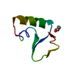
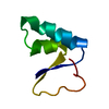
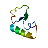
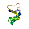
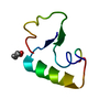
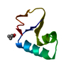
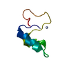
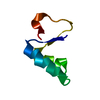
 Components Components | Crambin | ||||||
 Keywords Keywords | PLANT PROTEIN / thionin family | ||||||
| Function / homology |  Function and homology information Function and homology information | ||||||
| Biological species |  Crambe hispanica subsp. abyssinica (Abyssinian crambe) Crambe hispanica subsp. abyssinica (Abyssinian crambe) | ||||||
| Method |  X-RAY DIFFRACTION / MOLECULAR REPLACEMENT / Resolution: 0.85 Å X-RAY DIFFRACTION / MOLECULAR REPLACEMENT / Resolution: 0.85 Å | ||||||
 Authors Authors | Chen, J.C.-H. | ||||||
 Citation Citation | Journal: Acta Crystallogr.,Sect.F / Year: 2012 Title: Room-temperature ultrahigh-resolution time-of-flight neutron and X-ray diffraction studies of H/D-exchanged crambin. Authors: Chen, J.C. / Fisher, Z. / Kovalevsky, A.Y. / Mustyakimov, M. / Hanson, B.L. / Zhurov, V.V. / Langan, P. | ||||||
| History |
|
- Structure visualization
| Structure viewer | Molecule: MolmilJmol/JSmol |
|---|
- Downloads & links
-Download
| PDBx/mmCIF format | 3u7t.cif.gz | 40.4 KB | Display | PDBx/mmCIF format |
|---|---|---|---|---|
| PDB format | pdb3u7t.ent.gz | 28 KB | Display | PDB format |
| PDBx/mmJSON format | 3u7t.json.gz | Tree view | PDBx/mmJSON format | |
| Others |  Other downloads Other downloads |
-Validation report
| Arichive directory | https://data.pdbj.org/pub/pdb/validation_reports/u7/3u7tftp://data.pdbj.org/pub/pdb/validation_reports/u7/3u7t | HTTPS FTP |
|---|
-Related structure data
| Related structure data |  1crnS S: Starting model for refinement |
|---|---|
| Similar structure data |


-Links
 PDBj
PDBj
- Assembly
| Deposited unit | 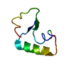
| ||||||||
|---|---|---|---|---|---|---|---|---|---|
| 1 |
| ||||||||
| Unit cell |
|
-Components
| #1: Protein/peptide | Mass: 4728.410 Da / Num. of mol.: 1 / Source method: isolated from a natural source Source: (natural) Crambe hispanica subsp. abyssinica (Abyssinian crambe)References: UniProt: P01542 |
|---|---|
| #2: Water | ChemComp-HOH /  Mass: 18.015 Da / Num. of mol.: 51 / Source method: isolated from a natural source / Formula: H2O Mass: 18.015 Da / Num. of mol.: 51 / Source method: isolated from a natural source / Formula: H2O |
| Has protein modification | Y |
-Experimental details
-Experiment
| Experiment | Method: X-RAY DIFFRACTION / Number of used crystals: 1 |
|---|
- Sample preparation
| Crystal | Density Matthews: 1.86 Å3/Da / Density % sol: 33.95 % |
|---|---|
| Crystal grow | Temperature: 298 K / Method: vapor diffusion, sitting drop / pH: 7 Details: Vapor diffusion of protein in 80 % EtOH solution against a 55 % EtOH solution, pH 7.0, VAPOR DIFFUSION, SITTING DROP, temperature 298K |
-Data collection
| Diffraction | Mean temperature: 298 K |
|---|---|
| Diffraction source | Source: ROTATING ANODE / Type: RIGAKU ULTRAX 18 / Wavelength: 0.71073 Å |
| Detector | Type: RIGAKU RAPID / Detector: IMAGE PLATE / Date: Nov 7, 2010 |
| Radiation | Monochromator: graphite / Protocol: SINGLE WAVELENGTH / Monochromatic (M) / Laue (L): M / Scattering type: x-ray |
| Radiation wavelength | Wavelength: 0.71073 Å / Relative weight: 1 |
| Reflection | Resolution: 0.85→30 Å / Num. all: 31025 / Num. obs: 30963 / % possible obs: 99.8 % / Observed criterion σ(F): 0 / Observed criterion σ(I): 0 / Redundancy: 3.41 % / Biso Wilson estimate: 6.3 Å2 / Rsym value: 0.05 / Net I/σ(I): 8.2 |
| Reflection shell | Resolution: 0.85→0.88 Å / Redundancy: 3.21 % / Rmerge(I) obs: 0.342 / Mean I/σ(I) obs: 1.9 / Num. unique all: 3065 / % possible all: 99.9 |
- Processing
| Software |
| ||||||||||||||||||||||||||||||||||||||||||||||||||||||||||||||||||||||||||||||||||||
|---|---|---|---|---|---|---|---|---|---|---|---|---|---|---|---|---|---|---|---|---|---|---|---|---|---|---|---|---|---|---|---|---|---|---|---|---|---|---|---|---|---|---|---|---|---|---|---|---|---|---|---|---|---|---|---|---|---|---|---|---|---|---|---|---|---|---|---|---|---|---|---|---|---|---|---|---|---|---|---|---|---|---|---|---|---|
| Refinement | Method to determine structure: MOLECULAR REPLACEMENT Starting model: PDB ENTRY 1CRN Resolution: 0.85→9.898 Å / SU ML: 0.07 / σ(F): 0 / Phase error: 12.19 / Stereochemistry target values: ML
| ||||||||||||||||||||||||||||||||||||||||||||||||||||||||||||||||||||||||||||||||||||
| Solvent computation | Shrinkage radii: 0.17 Å / VDW probe radii: 0.4 Å / Solvent model: FLAT BULK SOLVENT MODEL / Bsol: 80 Å2 / ksol: 0.6 e/Å3 | ||||||||||||||||||||||||||||||||||||||||||||||||||||||||||||||||||||||||||||||||||||
| Displacement parameters |
| ||||||||||||||||||||||||||||||||||||||||||||||||||||||||||||||||||||||||||||||||||||
| Refine analyze | Luzzati coordinate error obs: 0.07 Å | ||||||||||||||||||||||||||||||||||||||||||||||||||||||||||||||||||||||||||||||||||||
| Refinement step | Cycle: LAST / Resolution: 0.85→9.898 Å
| ||||||||||||||||||||||||||||||||||||||||||||||||||||||||||||||||||||||||||||||||||||
| Refine LS restraints |
| ||||||||||||||||||||||||||||||||||||||||||||||||||||||||||||||||||||||||||||||||||||
| LS refinement shell |
|
