 Movie
Movie Controller


+ Open data

- Basic information
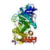
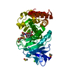
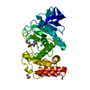
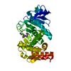
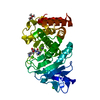
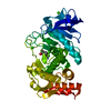
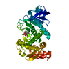
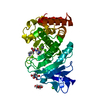
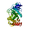
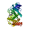
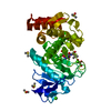
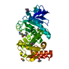
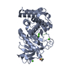
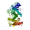
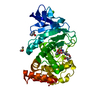
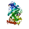
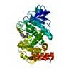
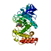
| Entry | Database: PDB / ID: 3msa | ||||||
|---|---|---|---|---|---|---|---|
| Title | Crystal structure of Thermolysin in complex with 3-Bromophenol | ||||||
 Components Components | Thermolysin | ||||||
 Keywords Keywords | HYDROLASE / PROTEASE / Fragment soaking / METALLOPROTEASE / Metal-binding / Secreted / Zymogen / 3-Bromophenol / Fragment based lead discovery | ||||||
| Function / homology |  Function and homology information Function and homology informationthermolysin / metalloendopeptidase activity / proteolysis / extracellular region / metal ion binding Similarity search - Function | ||||||
| Biological species |  | ||||||
| Method |  X-RAY DIFFRACTION / SYNCHROTRON / FOURIER SYNTHESIS / Resolution: 1.66 Å X-RAY DIFFRACTION / SYNCHROTRON / FOURIER SYNTHESIS / Resolution: 1.66 Å | ||||||
 Authors Authors | Behnen, J. / Heine, A. / Klebe, G. | ||||||
 Citation Citation | Journal: Chemmedchem / Year: 2012 Title: Experimental and computational active site mapping as a starting point to fragment-based lead discovery. Authors: Behnen, J. / Koster, H. / Neudert, G. / Craan, T. / Heine, A. / Klebe, G. | ||||||
| History |
|
- Structure visualization
| Structure viewer | Molecule: MolmilJmol/JSmol |
|---|
- Downloads & links
-Download
| PDBx/mmCIF format | 3msa.cif.gz | 83.4 KB | Display | PDBx/mmCIF format |
|---|---|---|---|---|
| PDB format | pdb3msa.ent.gz | 60.4 KB | Display | PDB format |
| PDBx/mmJSON format | 3msa.json.gz | Tree view | PDBx/mmJSON format | |
| Others |  Other downloads Other downloads |
-Validation report
| Arichive directory | https://data.pdbj.org/pub/pdb/validation_reports/ms/3msaftp://data.pdbj.org/pub/pdb/validation_reports/ms/3msa | HTTPS FTP |
|---|
-Related structure data
| Related structure data |  3ms3C  3msfC 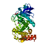 3msnC  3n21C 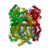 3n4aC 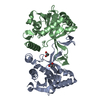 3n9wC  3nn7C 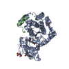 3nx8C 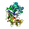 3pczC 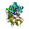 3prsC  3pvkC 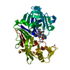 3pwwC  2a7gS C: citing same article ( S: Starting model for refinement |
|---|---|
| Similar structure data |


-Links
 PDBj
PDBj
- Assembly
| Deposited unit | 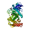
| |||||||||
|---|---|---|---|---|---|---|---|---|---|---|
| 1 |
| |||||||||
| Unit cell |
| |||||||||
| Components on special symmetry positions |
|
-Components
-Protein , 1 types, 1 molecules A
| #1: Protein | Mass: 34360.336 Da / Num. of mol.: 1 / Source method: isolated from a natural source / Source: (natural) |
|---|
-Non-polymers , 6 types, 229 molecules 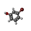


| #2: Chemical |  Mass: 173.007 Da / Num. of mol.: 2 / Source method: obtained synthetically / Formula: C6H5BrO Mass: 173.007 Da / Num. of mol.: 2 / Source method: obtained synthetically / Formula: C6H5BrO#3: Chemical | ChemComp-ZN / |  Mass: 65.409 Da / Num. of mol.: 1 / Source method: obtained synthetically / Formula: Zn Mass: 65.409 Da / Num. of mol.: 1 / Source method: obtained synthetically / Formula: Zn#4: Chemical | ChemComp-CA /  Mass: 40.078 Da / Num. of mol.: 4 / Source method: obtained synthetically / Formula: Ca Mass: 40.078 Da / Num. of mol.: 4 / Source method: obtained synthetically / Formula: Ca#5: Chemical |  Mass: 92.094 Da / Num. of mol.: 2 / Source method: obtained synthetically / Formula: C3H8O3 Mass: 92.094 Da / Num. of mol.: 2 / Source method: obtained synthetically / Formula: C3H8O3#6: Chemical | ChemComp-IPA / |  Mass: 60.095 Da / Num. of mol.: 1 / Source method: obtained synthetically / Formula: C3H8O Mass: 60.095 Da / Num. of mol.: 1 / Source method: obtained synthetically / Formula: C3H8O#7: Water | ChemComp-HOH / | Mass: 18.015 Da / Num. of mol.: 219 / Source method: isolated from a natural source / Formula: H2O |
|---|
-Experimental details
-Experiment
| Experiment | Method: X-RAY DIFFRACTION / Number of used crystals: 1 |
|---|
- Sample preparation
| Crystal | Density Matthews: 2.34 Å3/Da / Density % sol: 47.53 % |
|---|---|
| Crystal grow | Temperature: 288 K / Method: vapor diffusion, sitting drop / pH: 7.5 Details: 50 mM Tris/HCl, 50 % DMSO, 1.8 CsCl, pH 7.5, VAPOR DIFFUSION, SITTING DROP, temperature 288K |
-Data collection
| Diffraction | Mean temperature: 100 K |
|---|---|
| Diffraction source | Source: SYNCHROTRON / Site: BESSY  / Beamline: 14.2 / Wavelength: 0.91841 Å / Beamline: 14.2 / Wavelength: 0.91841 Å |
| Detector | Type: MARMOSAIC 225 mm CCD / Detector: CCD / Date: Feb 16, 2008 / Details: silicon |
| Radiation | Protocol: SINGLE WAVELENGTH / Monochromatic (M) / Laue (L): M / Scattering type: x-ray |
| Radiation wavelength | Wavelength: 0.91841 Å / Relative weight: 1 |
| Reflection | Resolution: 1.66→25 Å / Num. all: 36281 / Num. obs: 36281 / % possible obs: 94.9 % / Observed criterion σ(F): 0 / Observed criterion σ(I): 0 / Redundancy: 9.4 % / Rsym value: 0.083 / Net I/σ(I): 21.6 |
| Reflection shell | Resolution: 1.66→1.69 Å / Redundancy: 9.6 % / Mean I/σ(I) obs: 4.8 / Num. unique all: 1846 / Rsym value: 0.451 / % possible all: 94.4 |
- Processing
| Software |
| |||||||||||||||||||||||||||||||||
|---|---|---|---|---|---|---|---|---|---|---|---|---|---|---|---|---|---|---|---|---|---|---|---|---|---|---|---|---|---|---|---|---|---|---|
| Refinement | Method to determine structure: FOURIER SYNTHESIS Starting model: PDB entry 2A7G Resolution: 1.66→10 Å / Num. parameters: 10796 / Num. restraintsaints: 10319 / Cross valid method: FREE R / σ(F): 0 / Stereochemistry target values: ENGH AND HUBER
| |||||||||||||||||||||||||||||||||
| Refine analyze | Num. disordered residues: 5 / Occupancy sum hydrogen: 2251 / Occupancy sum non hydrogen: 2660.12 | |||||||||||||||||||||||||||||||||
| Refinement step | Cycle: LAST / Resolution: 1.66→10 Å
| |||||||||||||||||||||||||||||||||
| Refine LS restraints |
| |||||||||||||||||||||||||||||||||
| LS refinement shell | Resolution: 1.66→1.69 Å / Num. reflection obs: 36281 |
