 Movie
Movie Controller


+ Open data

- Basic information
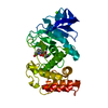
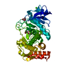
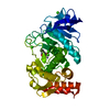
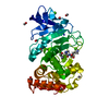
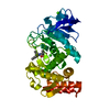
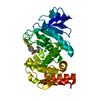
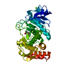
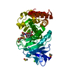
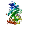
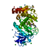
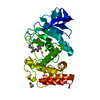
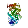
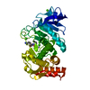
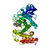
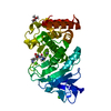
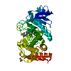
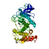
| Entry | Database: PDB / ID: 5wr5 | ||||||
|---|---|---|---|---|---|---|---|
| Title | Thermolysin, liganded form with cryo condition 1 | ||||||
 Components Components | Thermolysin | ||||||
 Keywords Keywords | HYDROLASE / X-ray free-electron laser / Serial femtosecond crystallography / Microcrystal / Structure-based drug design | ||||||
| Function / homology |  Function and homology information Function and homology informationthermolysin / metalloendopeptidase activity / proteolysis / extracellular region / metal ion binding Similarity search - Function | ||||||
| Biological species |   Geobacillus stearothermophilus (bacteria) Geobacillus stearothermophilus (bacteria) | ||||||
| Method |  X-RAY DIFFRACTION / SYNCHROTRON / MOLECULAR REPLACEMENT / Resolution: 1.9 Å X-RAY DIFFRACTION / SYNCHROTRON / MOLECULAR REPLACEMENT / Resolution: 1.9 Å | ||||||
 Authors Authors | Kunishima, N. / Naitow, H. / Matsuura, Y. | ||||||
 Citation Citation | Journal: Acta Crystallogr D Struct Biol / Year: 2017 Title: Protein-ligand complex structure from serial femtosecond crystallography using soaked thermolysin microcrystals and comparison with structures from synchrotron radiation Authors: Naitow, H. / Matsuura, Y. / Tono, K. / Joti, Y. / Kameshima, T. / Hatsui, T. / Yabashi, M. / Tanaka, R. / Tanaka, T. / Sugahara, M. / Kobayashi, J. / Nango, E. / Iwata, S. / Kunishima, N. | ||||||
| History |
|
- Structure visualization
| Structure viewer | Molecule: MolmilJmol/JSmol |
|---|
- Downloads & links
-Download
| PDBx/mmCIF format | 5wr5.cif.gz | 88.1 KB | Display | PDBx/mmCIF format |
|---|---|---|---|---|
| PDB format | pdb5wr5.ent.gz | 64.2 KB | Display | PDB format |
| PDBx/mmJSON format | 5wr5.json.gz | Tree view | PDBx/mmJSON format | |
| Others |  Other downloads Other downloads |
-Validation report
| Arichive directory | https://data.pdbj.org/pub/pdb/validation_reports/wr/5wr5ftp://data.pdbj.org/pub/pdb/validation_reports/wr/5wr5 | HTTPS FTP |
|---|
-Related structure data
| Related structure data | 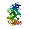 5wr2C  5wr3C  5wr4C  5wr6C 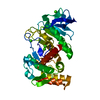 4ow3S C: citing same article ( S: Starting model for refinement |
|---|---|
| Similar structure data |


-Links
 PDBj
PDBj- Assembly
| Deposited unit | 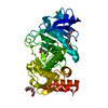
| |||||||||
|---|---|---|---|---|---|---|---|---|---|---|
| 1 |
| |||||||||
| Unit cell |
| |||||||||
| Components on special symmetry positions |
|
-Components
-Protein , 1 types, 1 molecules A
| #1: Protein | Mass: 34360.336 Da / Num. of mol.: 1 / Fragment: UNP residues 236-551 Source method: isolated from a genetically manipulated source Source: (gene. exp.) Geobacillus stearothermophilus (bacteria)Gene: nprS, nprM / Production host: Geobacillus stearothermophilus (bacteria) / References: UniProt: P43133, thermolysin |
|---|
-Non-polymers , 5 types, 392 molecules 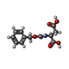


| #2: Chemical | ChemComp-NX6 /  Mass: 267.235 Da / Num. of mol.: 1 / Source method: obtained synthetically / Formula: C12H13NO6 Mass: 267.235 Da / Num. of mol.: 1 / Source method: obtained synthetically / Formula: C12H13NO6 | ||||
|---|---|---|---|---|---|
| #3: Chemical | ChemComp-ZN /  Mass: 65.409 Da / Num. of mol.: 1 / Source method: obtained synthetically / Formula: Zn Mass: 65.409 Da / Num. of mol.: 1 / Source method: obtained synthetically / Formula: Zn | ||||
| #4: Chemical | ChemComp-CA /  Mass: 40.078 Da / Num. of mol.: 4 / Source method: obtained synthetically / Formula: Ca Mass: 40.078 Da / Num. of mol.: 4 / Source method: obtained synthetically / Formula: Ca#5: Chemical |  Mass: 194.226 Da / Num. of mol.: 2 / Source method: obtained synthetically / Formula: C8H18O5 / Comment: precipitant*YM Mass: 194.226 Da / Num. of mol.: 2 / Source method: obtained synthetically / Formula: C8H18O5 / Comment: precipitant*YM#6: Water | ChemComp-HOH / | Mass: 18.015 Da / Num. of mol.: 384 / Source method: isolated from a natural source / Formula: H2O |
-Experimental details
-Experiment
| Experiment | Method: X-RAY DIFFRACTION / Number of used crystals: 1 |
|---|
- Sample preparation
| Crystal | Density Matthews: 2.32 Å3/Da / Density % sol: 46.95 % |
|---|---|
| Crystal grow | Temperature: 293 K / Method: vapor diffusion, hanging drop / pH: 6.5 / Details: PEG2000MME, MES, CaCl2 |
-Data collection
| Diffraction | Mean temperature: 100 K |
|---|---|
| Diffraction source | Source: SYNCHROTRON / Site: SPring-8  / Beamline: BL26B2 / Wavelength: 1 Å / Beamline: BL26B2 / Wavelength: 1 Å |
| Detector | Type: MARMOSAIC 225 mm CCD / Detector: CCD / Date: May 13, 2015 |
| Radiation | Protocol: SINGLE WAVELENGTH / Monochromatic (M) / Laue (L): M / Scattering type: x-ray |
| Radiation wavelength | Wavelength: 1 Å / Relative weight: 1 |
| Reflection | Resolution: 1.9→46.1 Å / Num. obs: 26366 / % possible obs: 100 % / Redundancy: 8.6 % / Biso Wilson estimate: 20.56 Å2 / Rmerge(I) obs: 0.086 / Net I/σ(I): 21.1 |
| Reflection shell | Resolution: 1.9→1.97 Å / Redundancy: 6.5 % / Rmerge(I) obs: 0.503 / Mean I/σ(I) obs: 3.8 / % possible all: 100 |
- Processing
| Software |
| ||||||||||||||||||||
|---|---|---|---|---|---|---|---|---|---|---|---|---|---|---|---|---|---|---|---|---|---|
| Refinement | Method to determine structure: MOLECULAR REPLACEMENT Starting model: 4OW3 Resolution: 1.9→43.5 Å / SU ML: 0.17 / Cross valid method: THROUGHOUT / Phase error: 16.99
| ||||||||||||||||||||
| Displacement parameters | Biso mean: 22.79 Å2 | ||||||||||||||||||||
| Refinement step | Cycle: LAST / Resolution: 1.9→43.5 Å
| ||||||||||||||||||||
| LS refinement shell | Resolution: 1.9→1.98 Å
|
