 Movie
Movie Controller

[English] 日本語
 Yorodumi
- PDB-1gxw: the 2.2 A resolution structure of thermolysin crystallized in pre... -
+ Open data

- Basic information
| Entry | Database: PDB / ID: 1gxw | ||||||
|---|---|---|---|---|---|---|---|
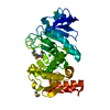
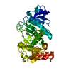
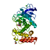
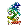
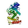
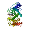
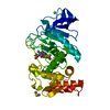
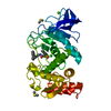
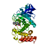
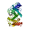
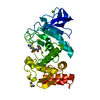
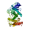
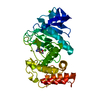
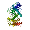
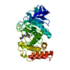
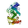
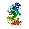
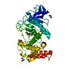
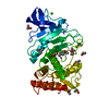
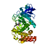
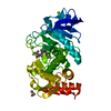
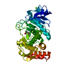
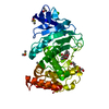
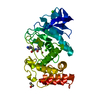
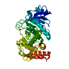
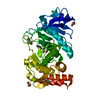
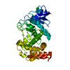
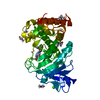
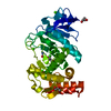
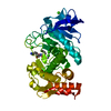
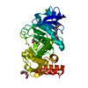
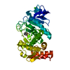
| Title | the 2.2 A resolution structure of thermolysin crystallized in presence of potassium thiocyanate | ||||||
 Components Components | THERMOLYSIN | ||||||
 Keywords Keywords | HYDROLASE / THERMOLYSIN / METALLOENDOPEPTIDASE / THIOCYANATE / SALTING-IN / METALLOPROTEASE | ||||||
| Function / homology |  Function and homology information Function and homology informationthermolysin / metalloendopeptidase activity / proteolysis / extracellular region / metal ion binding Similarity search - Function | ||||||
| Biological species |  | ||||||
| Method |  X-RAY DIFFRACTION / SYNCHROTRON / MOLECULAR REPLACEMENT / Resolution: 2.18 Å X-RAY DIFFRACTION / SYNCHROTRON / MOLECULAR REPLACEMENT / Resolution: 2.18 Å | ||||||
 Authors Authors | Gaucher, J.F. / Selkti, M. / Prange, T. / Tomas, A. | ||||||
 Citation Citation | Journal: Acta Crystallogr.,Sect.D / Year: 2002 Title: The 2.2 A Resolution Structure of Thermolysin (Tln) Crystallized in the Presence of Potassium Thiocyanate. Authors: Gaucher, J. / Selkti, M. / Prange, T. / Tomas, A. #1: Journal: J.Biochem. / Year: 1998 Title: Effect of Salts on the Solubility of Thermolysin: A Remarkable Increase in the Solubility as Well as the Activity by the Addition of Salts without Aggregation or Dispersion of Thermolysin Authors: Inouye, K. / Kuzuya, K. / Tonomura, B. #2: Journal: J.Mol.Biol. / Year: 1982 Title: Structure of Thermolysin Refined at 1.6A Resolution Authors: Holmes, M.A. / Matthews, B.W. | ||||||
| History |
| ||||||
| Remark 700 | SHEET THE SHEET STRUCTURE OF THIS MOLECULE IS BIFURCATED. IN ORDER TO REPRESENT THIS FEATURE IN ... SHEET THE SHEET STRUCTURE OF THIS MOLECULE IS BIFURCATED. IN ORDER TO REPRESENT THIS FEATURE IN THE SHEET RECORDS BELOW, TWO SHEETS ARE DEFINED. |
- Structure visualization
| Structure viewer | Molecule: MolmilJmol/JSmol |
|---|
- Downloads & links
-Download
| PDBx/mmCIF format | 1gxw.cif.gz | 81.6 KB | Display | PDBx/mmCIF format |
|---|---|---|---|---|
| PDB format | pdb1gxw.ent.gz | 60.2 KB | Display | PDB format |
| PDBx/mmJSON format | 1gxw.json.gz | Tree view | PDBx/mmJSON format | |
| Others |  Other downloads Other downloads |
-Validation report
| Arichive directory | https://data.pdbj.org/pub/pdb/validation_reports/gx/1gxwftp://data.pdbj.org/pub/pdb/validation_reports/gx/1gxw | HTTPS FTP |
|---|
-Related structure data
| Related structure data |  8tlnS S: Starting model for refinement |
|---|---|
| Similar structure data |


-Links
 PDBj
PDBj

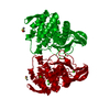
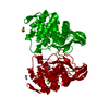
- Assembly
| Deposited unit | 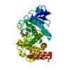
| ||||||||
|---|---|---|---|---|---|---|---|---|---|
| 1 |
| ||||||||
| Unit cell |
| ||||||||
| Details | THE BIOMOLECULE CONSISTS OF A DI-PEPTIDE BURIED WITHINTHE PROTEIN. THE BOUND SURFACE AREA BETWEEN THE TWOCHAINS IS 947.8 ANGSTROM**2 |
-Components
-Protein , 1 types, 1 molecules A
| #1: Protein | Mass: 34362.305 Da / Num. of mol.: 1 / Source method: isolated from a natural source / Source: (natural) |
|---|
-Non-polymers , 6 types, 178 molecules 


| #2: Chemical | ChemComp-VAL /  Type: L-peptide linking / Mass: 117.146 Da / Num. of mol.: 1 / Source method: obtained synthetically / Formula: C5H11NO2 Type: L-peptide linking / Mass: 117.146 Da / Num. of mol.: 1 / Source method: obtained synthetically / Formula: C5H11NO2 | ||||||
|---|---|---|---|---|---|---|---|
| #3: Chemical | ChemComp-LYS /  Type: L-peptide linking / Mass: 147.195 Da / Num. of mol.: 1 / Source method: obtained synthetically / Formula: C6H15N2O2 Type: L-peptide linking / Mass: 147.195 Da / Num. of mol.: 1 / Source method: obtained synthetically / Formula: C6H15N2O2 | ||||||
| #4: Chemical | ChemComp-CA /  Mass: 40.078 Da / Num. of mol.: 4 / Source method: obtained synthetically / Formula: Ca Mass: 40.078 Da / Num. of mol.: 4 / Source method: obtained synthetically / Formula: Ca#5: Chemical | ChemComp-ZN / |  Mass: 65.409 Da / Num. of mol.: 1 / Source method: obtained synthetically / Formula: Zn Mass: 65.409 Da / Num. of mol.: 1 / Source method: obtained synthetically / Formula: Zn#6: Chemical | ChemComp-SCN / |  Mass: 58.082 Da / Num. of mol.: 1 / Source method: obtained synthetically / Formula: CNS Mass: 58.082 Da / Num. of mol.: 1 / Source method: obtained synthetically / Formula: CNS#7: Water | ChemComp-HOH / | Mass: 18.015 Da / Num. of mol.: 170 / Source method: isolated from a natural source / Formula: H2O |
-Experimental details
-Experiment
| Experiment | Method: X-RAY DIFFRACTION / Number of used crystals: 1 |
|---|
- Sample preparation
| Crystal | Density Matthews: 2.38 Å3/Da / Density % sol: 38.8 % | |||||||||||||||||||||||||||||||||||
|---|---|---|---|---|---|---|---|---|---|---|---|---|---|---|---|---|---|---|---|---|---|---|---|---|---|---|---|---|---|---|---|---|---|---|---|---|
| Crystal grow | Method: vapor diffusion, hanging drop / pH: 8.3 Details: 0.5MICROL HANGING DROP CONTAINING 87MG/ML OF THERMOLYSIN IN BUFFER A (50MM TRIS/HCL PH8.3, 1M KSCN, 20MM CACL2) WAS EQUILIBRATED AGAINST 740MICROL BUFFER A AND 260MICROL WATER (SALTING-IN METHOD), pH 8.30 | |||||||||||||||||||||||||||||||||||
| Crystal grow | *PLUS Temperature: 277 K / Method: vapor diffusion | |||||||||||||||||||||||||||||||||||
| Components of the solutions | *PLUS
|
-Data collection
| Diffraction | Mean temperature: 283 K |
|---|---|
| Diffraction source | Source: SYNCHROTRON / Site: LURE  / Beamline: DW21B / Wavelength: 1.375 / Beamline: DW21B / Wavelength: 1.375 |
| Detector | Type: MARRESEARCH / Detector: IMAGE PLATE / Date: Jul 15, 1999 / Details: BENT MIRROR |
| Radiation | Monochromator: SI(111) / Protocol: SINGLE WAVELENGTH / Monochromatic (M) / Laue (L): M / Scattering type: x-ray |
| Radiation wavelength | Wavelength: 1.375 Å / Relative weight: 1 |
| Reflection | Resolution: 2.18→19 Å / Num. obs: 15898 / % possible obs: 92.4 % / Redundancy: 3.7 % / Biso Wilson estimate: 12.1 Å2 / Rmerge(I) obs: 0.086 / Net I/σ(I): 7.3 |
| Reflection shell | Resolution: 2.18→2.32 Å / % possible all: 83.9 |
| Reflection | *PLUS Lowest resolution: 18.5 Å / % possible obs: 92.2 % / Num. measured all: 58898 |
| Reflection shell | *PLUS Lowest resolution: 2.3 Å / % possible obs: 82.7 % / Rmerge(I) obs: 0.241 / Mean I/σ(I) obs: 2.3 |
- Processing
| Software |
| ||||||||||||||||||||||||||||||||||||||||||||||||||||||||||||||||||||||||||||||||
|---|---|---|---|---|---|---|---|---|---|---|---|---|---|---|---|---|---|---|---|---|---|---|---|---|---|---|---|---|---|---|---|---|---|---|---|---|---|---|---|---|---|---|---|---|---|---|---|---|---|---|---|---|---|---|---|---|---|---|---|---|---|---|---|---|---|---|---|---|---|---|---|---|---|---|---|---|---|---|---|---|---|
| Refinement | Method to determine structure: MOLECULAR REPLACEMENT Starting model: PDB ENTRY 8TLN Resolution: 2.18→18.46 Å / Rfactor Rfree error: 0.005 / Data cutoff high absF: 2790416.67 / Isotropic thermal model: RESTRAINED / Cross valid method: THROUGHOUT / σ(F): 0 / Details: BULK SOLVENT MODEL USED
| ||||||||||||||||||||||||||||||||||||||||||||||||||||||||||||||||||||||||||||||||
| Solvent computation | Solvent model: FLAT MODEL / Bsol: 105.015 Å2 / ksol: 0.590377 e/Å3 | ||||||||||||||||||||||||||||||||||||||||||||||||||||||||||||||||||||||||||||||||
| Displacement parameters | Biso mean: 16.6 Å2
| ||||||||||||||||||||||||||||||||||||||||||||||||||||||||||||||||||||||||||||||||
| Refine analyze |
| ||||||||||||||||||||||||||||||||||||||||||||||||||||||||||||||||||||||||||||||||
| Refinement step | Cycle: LAST / Resolution: 2.18→18.46 Å
| ||||||||||||||||||||||||||||||||||||||||||||||||||||||||||||||||||||||||||||||||
| Refine LS restraints |
| ||||||||||||||||||||||||||||||||||||||||||||||||||||||||||||||||||||||||||||||||
| LS refinement shell | Resolution: 2.18→2.32 Å / Rfactor Rfree error: 0.015 / Total num. of bins used: 6
| ||||||||||||||||||||||||||||||||||||||||||||||||||||||||||||||||||||||||||||||||
| Xplor file |
| ||||||||||||||||||||||||||||||||||||||||||||||||||||||||||||||||||||||||||||||||
| Refinement | *PLUS Lowest resolution: 19 Å / Rfactor Rfree: 0.214 | ||||||||||||||||||||||||||||||||||||||||||||||||||||||||||||||||||||||||||||||||
| Solvent computation | *PLUS | ||||||||||||||||||||||||||||||||||||||||||||||||||||||||||||||||||||||||||||||||
| Displacement parameters | *PLUS | ||||||||||||||||||||||||||||||||||||||||||||||||||||||||||||||||||||||||||||||||
| Refine LS restraints | *PLUS
| ||||||||||||||||||||||||||||||||||||||||||||||||||||||||||||||||||||||||||||||||
| LS refinement shell | *PLUS Rfactor Rfree: 0.242 |
