 Movie
Movie Controller


+ Open data

- Basic information
| Entry | Database: PDB / ID: 1fjt | ||||||
|---|---|---|---|---|---|---|---|
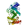
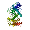
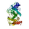
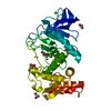
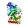
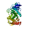
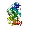
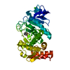
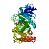
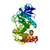
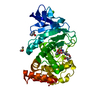
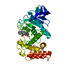
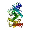
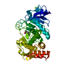
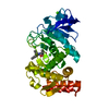
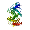
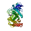
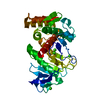
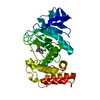
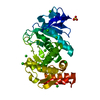
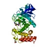
| Title | THERMOLYSIN (50% ACETONITRILE SOAKED CRYSTALS) | ||||||
 Components Components | THERMOLYSIN | ||||||
 Keywords Keywords | HYDROLASE / METALLOPROTEINASE / ORGANIC SOLVENT | ||||||
| Function / homology |  Function and homology information Function and homology informationthermolysin / metalloendopeptidase activity / proteolysis / extracellular region / metal ion binding Similarity search - Function | ||||||
| Biological species |  | ||||||
| Method |  X-RAY DIFFRACTION / isomorphous replacement / Resolution: 2.2 Å X-RAY DIFFRACTION / isomorphous replacement / Resolution: 2.2 Å | ||||||
 Authors Authors | English, A.C. / Groom, C.R. / Hubbard, R.E. | ||||||
 Citation Citation | Journal: Protein Eng. / Year: 2001 Title: Experimental and computational mapping of the binding surface of a crystalline protein. Authors: English, A.C. / Groom, C.R. / Hubbard, R.E. | ||||||
| History |
|
- Structure visualization
| Structure viewer | Molecule: MolmilJmol/JSmol |
|---|
- Downloads & links
-Download
| PDBx/mmCIF format | 1fjt.cif.gz | 79.8 KB | Display | PDBx/mmCIF format |
|---|---|---|---|---|
| PDB format | pdb1fjt.ent.gz | 59.8 KB | Display | PDB format |
| PDBx/mmJSON format | 1fjt.json.gz | Tree view | PDBx/mmJSON format | |
| Others |  Other downloads Other downloads |
-Validation report
| Arichive directory | https://data.pdbj.org/pub/pdb/validation_reports/fj/1fjtftp://data.pdbj.org/pub/pdb/validation_reports/fj/1fjt | HTTPS FTP |
|---|
-Related structure data
| Related structure data |  1fj3C  1fjoC  1fjqC  1fjuC  1fjvC 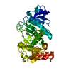 1fjwC C: citing same article ( |
|---|---|
| Similar structure data |


-Links
 PDBj
PDBj
- Assembly
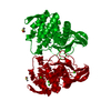
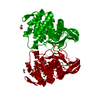
| Deposited unit | 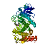
| ||||||||
|---|---|---|---|---|---|---|---|---|---|
| 1 |
| ||||||||
| Unit cell |
|
-Components
-Protein , 1 types, 1 molecules A
| #1: Protein | Mass: 34362.305 Da / Num. of mol.: 1 / Source method: isolated from a natural source / Source: (natural) |
|---|
-Non-polymers , 6 types, 167 molecules 


| #2: Chemical | ChemComp-ZN /  Mass: 65.409 Da / Num. of mol.: 1 / Source method: obtained synthetically / Formula: Zn Mass: 65.409 Da / Num. of mol.: 1 / Source method: obtained synthetically / Formula: Zn | ||||||||
|---|---|---|---|---|---|---|---|---|---|
| #3: Chemical | ChemComp-CA /  Mass: 40.078 Da / Num. of mol.: 4 / Source method: obtained synthetically / Formula: Ca Mass: 40.078 Da / Num. of mol.: 4 / Source method: obtained synthetically / Formula: Ca#4: Chemical | ChemComp-DMS / |  Mass: 78.133 Da / Num. of mol.: 1 / Source method: obtained synthetically / Formula: C2H6OS / Comment: DMSO, precipitant*YM Mass: 78.133 Da / Num. of mol.: 1 / Source method: obtained synthetically / Formula: C2H6OS / Comment: DMSO, precipitant*YM#5: Chemical | ChemComp-VAL / |  Type: L-peptide linking / Mass: 117.146 Da / Num. of mol.: 1 / Source method: obtained synthetically / Formula: C5H11NO2 Type: L-peptide linking / Mass: 117.146 Da / Num. of mol.: 1 / Source method: obtained synthetically / Formula: C5H11NO2#6: Chemical | ChemComp-LYS / |  Type: L-peptide linking / Mass: 147.195 Da / Num. of mol.: 1 / Source method: obtained synthetically / Formula: C6H15N2O2 Type: L-peptide linking / Mass: 147.195 Da / Num. of mol.: 1 / Source method: obtained synthetically / Formula: C6H15N2O2#7: Water | ChemComp-HOH / | Mass: 18.015 Da / Num. of mol.: 159 / Source method: isolated from a natural source / Formula: H2O |
-Experimental details
-Experiment
| Experiment | Method: X-RAY DIFFRACTION / Number of used crystals: 1 |
|---|
- Sample preparation
| Crystal | Density Matthews: 2.42 Å3/Da / Density % sol: 49.23 % | ||||||||||||||||||||||||||||||
|---|---|---|---|---|---|---|---|---|---|---|---|---|---|---|---|---|---|---|---|---|---|---|---|---|---|---|---|---|---|---|---|
| Crystal grow | Temperature: 298 K / Method: vapor diffusion, sitting drop / pH: 7.5 Details: 4.6MM THERMOLYSIN, 45% DMSO, 50MM TRIS-HCL, 2.5M CSCL, pH 7.5, VAPOR DIFFUSION, SITTING DROP, temperature 298K | ||||||||||||||||||||||||||||||
| Crystal grow | *PLUS Method: unknownDetails: English, A.C., (1999) Proteins Struct.Funct.Genet., 37, 628. | ||||||||||||||||||||||||||||||
| Components of the solutions | *PLUS
|
-Data collection
| Diffraction | Mean temperature: 298 K |
|---|---|
| Diffraction source | Source: ROTATING ANODE / Type: RIGAKU / Wavelength: 1.5418 |
| Detector | Type: MARRESEARCH / Detector: AREA DETECTOR / Date: Nov 6, 2000 |
| Radiation | Protocol: SINGLE WAVELENGTH / Monochromatic (M) / Laue (L): M / Scattering type: x-ray |
| Radiation wavelength | Wavelength: 1.5418 Å / Relative weight: 1 |
| Reflection | Resolution: 2.2→20 Å / Num. all: 161170 / Num. obs: 19030 / % possible obs: 91 % / Observed criterion σ(I): 2 / Redundancy: 8.5 % / Biso Wilson estimate: 24 Å2 / Rmerge(I) obs: 0.09 / Net I/σ(I): 5.7 |
| Reflection shell | Resolution: 2.15→2.27 Å / Redundancy: 8.4 % / Rmerge(I) obs: 0.38 / Num. unique all: 22857 / % possible all: 91 |
| Reflection | *PLUS % possible obs: 93 % |
| Reflection shell | *PLUS % possible obs: 91 % / Mean I/σ(I) obs: 2 |
- Processing
| Software |
| ||||||||||||||||||||
|---|---|---|---|---|---|---|---|---|---|---|---|---|---|---|---|---|---|---|---|---|---|
| Refinement | Method to determine structure: isomorphous replacement / Resolution: 2.2→20 Å / σ(I): 2 / Stereochemistry target values: Engh & Huber Details: Maximim likelihood procedure used AT THIS CONCENTRATION (50% ACETONITRILE) THERE IS ELECTRON DENSITY CORRESPONDING TO THE DIPEPTIDE (VAL506-LYS507) OBSERVED IN THE ACTIVE SITE OF THE NATIVE ENZYME.
| ||||||||||||||||||||
| Refinement step | Cycle: LAST / Resolution: 2.2→20 Å
| ||||||||||||||||||||
| Refine LS restraints |
|
