 Movie
Movie Controller


+ Open data

- Basic information
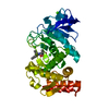
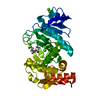
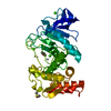
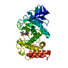
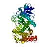
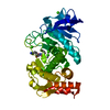
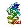
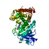
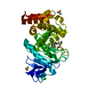
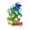
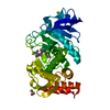
| Entry | Database: PDB / ID: 1lna | ||||||
|---|---|---|---|---|---|---|---|
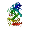
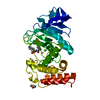
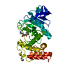
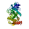
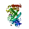
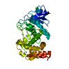
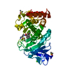
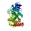
| Title | A STRUCTURAL ANALYSIS OF METAL SUBSTITUTIONS IN THERMOLYSIN | ||||||
 Components Components | THERMOLYSIN | ||||||
 Keywords Keywords | METALLOPROTEASE | ||||||
| Function / homology |  Function and homology information Function and homology informationthermolysin / metalloendopeptidase activity / proteolysis / extracellular region / metal ion binding Similarity search - Function | ||||||
| Biological species |  | ||||||
| Method |  X-RAY DIFFRACTION / MOLECULAR SUBSTITUTION / Resolution: 1.9 Å X-RAY DIFFRACTION / MOLECULAR SUBSTITUTION / Resolution: 1.9 Å | ||||||
 Authors Authors | Holland, D.R. / Hausrath, A.C. / Juers, D. / Matthews, B.W. | ||||||
 Citation Citation | Journal: Protein Sci. / Year: 1995 Title: Structural analysis of zinc substitutions in the active site of thermolysin. Authors: Holland, D.R. / Hausrath, A.C. / Juers, D. / Matthews, B.W. #1: Journal: Biochemistry / Year: 1992Title: Structural Comparison Suggests that Thermolysin and Related Neutral Proteases Undergo Hinge-Bending Motion During Catalysis Authors: Holland, D.R. / Tronrud, D.E. / Pley, H.W. / Flaherty, K.M. / Stark, W. / Jansonius, J.N. / Mckay, D.B. / Matthews, B.W. #2: Journal: J.Mol.Biol. / Year: 1982Title: Structure of Thermolysin Refined at 1.6 Angstroms Resolution Authors: Holmes, M.A. / Matthews, B.W. #3: Journal: Biochemistry / Year: 1982Title: Structure of a Mercaptan-Thermolysin Complex Illustrates Mode of Inhibition of Zinc Proteases by Substrate-Analogue Mercaptans Authors: Monzingo, A.F. / Matthews, B.W. #4: Journal: Biochemistry / Year: 1981Title: Binding of Hydroxamic Acid Inhibitors to Crystalline Thermolysin Suggests a Pentacoordinate Zinc Intermediate in Catalysis Authors: Holmes, M.A. / Matthews, B.W. #5: Journal: J.Biol.Chem. / Year: 1979Title: Binding of the Biproduct Analog L-Benzylsuccinic Acid to Thermolysin Determined by X-Ray Crystallography Authors: Bolognesi, M.C. / Matthews, B.W. #6: Journal: J.Biol.Chem. / Year: 1977Title: Comparison of the Structures of Carboxypeptidase a and Thermolysin Authors: Kester, W.R. / Matthews, B.W. #7: Journal: J.Mol.Biol. / Year: 1977Title: A Crystallographic Study of the Complex of Phosphoramidon with Thermolysin. A Model for the Presumed Catalytic Transition State and for the Binding of Extended Substrates Authors: Weaver, L.H. / Kester, W.R. / Matthews, B.W. #8: Journal: Biochemistry / Year: 1977Title: Crystallographic Study of the Binding of Dipeptide Inhibitors to Thermolysin. Implications for the Mechanism of Catalysis Authors: Kester, W.R. / Matthews, B.W. #9: Journal: Biochemistry / Year: 1976Title: Role of Calcium in the Thermal Stability of Thermolysin Authors: Dahlquist, F.W. / Long, J.W. / Bigbee, W.L. #11: Journal: Proc.Natl.Acad.Sci.USA / Year: 1975Title: Evidence of Homologous Relationship between Thermolysin and Neutral Protease a of Bacillus Subtilis Authors: Levy, P.L. / Pangburn, M.K. / Burstein, Y. / Ericsson, L.H. / Neurath, H. / Walsh, K.A. #12: Journal: J.Biol.Chem. / Year: 1974Title: The Conformation of Thermolysin Authors: Matthews, B.W. / Weaver, L.H. / Kester, W.R. #13: Journal: Biochemistry / Year: 1974Title: Binding of Lanthanide Ions to Thermolysin Authors: Matthews, B.W. / Weaver, L.H. #14: Journal: J.Mol.Biol. / Year: 1972Title: The Structure of Thermolysin,an Electron Density Map at 2.3 Angstroms Resolution Authors: Colman, P.M. / Jansonius, J.N. / Matthews, B.W. #15: Journal: Nature New Biol. / Year: 1972Title: Amino-Acid Sequence of Thermolysin Authors: Titani, K. / Hermodson, M.A. / Ericsson, L.H. / Walsh, K.A. / Neurath, H. #16: Journal: Nature New Biol. / Year: 1972Title: Three Dimensional Structure of Thermolysin Authors: Matthews, B.W. / Jansonius, J.N. / Colman, P.M. / Schoenborn, B.P. / Duporque, D. #17: Journal: Nature New Biol. / Year: 1972Title: Structure of Thermolysin Authors: Matthews, B.W. / Colman, P.M. / Jansonius, J.N. / Titani, K. / Walsh, K.A. / Neurath, H. | ||||||
| History |
|
- Structure visualization
| Structure viewer | Molecule: MolmilJmol/JSmol |
|---|
- Downloads & links
-Download
| PDBx/mmCIF format | 1lna.cif.gz | 81.3 KB | Display | PDBx/mmCIF format |
|---|---|---|---|---|
| PDB format | pdb1lna.ent.gz | 60.5 KB | Display | PDB format |
| PDBx/mmJSON format | 1lna.json.gz | Tree view | PDBx/mmJSON format | |
| Others |  Other downloads Other downloads |
-Validation report
| Arichive directory | https://data.pdbj.org/pub/pdb/validation_reports/ln/1lnaftp://data.pdbj.org/pub/pdb/validation_reports/ln/1lna | HTTPS FTP |
|---|
-Related structure data


-Links
 PDBj
PDBj

- Assembly
| Deposited unit | 
| ||||||||
|---|---|---|---|---|---|---|---|---|---|
| 1 |
| ||||||||
| Unit cell |
| ||||||||
| Atom site foot note | 1: CIS PROLINE - PRO E 51 |
-Components
-Protein , 1 types, 1 molecules E
| #1: Protein | Mass: 34362.305 Da / Num. of mol.: 1 Source method: isolated from a genetically manipulated source Source: (gene. exp.) References: UniProt: P00800, thermolysin |
|---|
-Non-polymers , 6 types, 166 molecules 


| #2: Chemical | ChemComp-VAL /  Type: L-peptide linking / Mass: 117.146 Da / Num. of mol.: 1 / Source method: obtained synthetically / Formula: C5H11NO2 Type: L-peptide linking / Mass: 117.146 Da / Num. of mol.: 1 / Source method: obtained synthetically / Formula: C5H11NO2 | ||||||
|---|---|---|---|---|---|---|---|
| #3: Chemical | ChemComp-LYS /  Type: L-peptide linking / Mass: 147.195 Da / Num. of mol.: 1 / Source method: obtained synthetically / Formula: C6H15N2O2 Type: L-peptide linking / Mass: 147.195 Da / Num. of mol.: 1 / Source method: obtained synthetically / Formula: C6H15N2O2 | ||||||
| #4: Chemical |  Mass: 58.933 Da / Num. of mol.: 2 / Source method: obtained synthetically / Formula: Co Mass: 58.933 Da / Num. of mol.: 2 / Source method: obtained synthetically / Formula: Co#5: Chemical |  Mass: 40.078 Da / Num. of mol.: 3 / Source method: obtained synthetically / Formula: Ca Mass: 40.078 Da / Num. of mol.: 3 / Source method: obtained synthetically / Formula: Ca#6: Chemical | ChemComp-DMS / |  Mass: 78.133 Da / Num. of mol.: 1 / Source method: obtained synthetically / Formula: C2H6OS / Comment: DMSO, precipitant*YM Mass: 78.133 Da / Num. of mol.: 1 / Source method: obtained synthetically / Formula: C2H6OS / Comment: DMSO, precipitant*YM#7: Water | ChemComp-HOH / | Mass: 18.015 Da / Num. of mol.: 158 / Source method: isolated from a natural source / Formula: H2O |
-Details
| Nonpolymer details | RESIDUES 1321 AND 1322 FORM A DIPEPTIDE (VAL-LYS) BOUND IN THE ACTIVE SITE OF THE MOLECULE. IT IS ...RESIDUES 1321 AND 1322 FORM A DIPEPTIDE (VAL-LYS) BOUND IN THE ACTIVE SITE OF THE MOLECULE. IT IS PRESUMED THAT THE ORIGIN OF THIS DIPEPTIDE IS THE C-TERMINAL TWO RESIDUES OF THE PROTEIN. SINCE THE C-TERMINUS APPEARS AT FULL OCCUPANCY, MOLECULES NOT INCORPORAT |
|---|
-Experimental details
-Experiment
| Experiment | Method: X-RAY DIFFRACTION |
|---|
- Sample preparation
| Crystal | Density Matthews: 2.41 Å3/Da / Density % sol: 48.88 % | ||||||||||||||||||||||||||||||
|---|---|---|---|---|---|---|---|---|---|---|---|---|---|---|---|---|---|---|---|---|---|---|---|---|---|---|---|---|---|---|---|
| Crystal grow | pH: 7.2 / Details: pH 7.2 | ||||||||||||||||||||||||||||||
| Crystal grow | *PLUS Method: unknown / Details: Holmes, M.A., (1982) J.Mol.Biol., 160, 623. | ||||||||||||||||||||||||||||||
| Components of the solutions | *PLUS
|
-Data collection
| Diffraction | Mean temperature: 298 K |
|---|---|
| Diffraction source | Source: ROTATING ANODE / Type: RIGAKU RU200 / Wavelength: 1.5418 |
| Detector | Type: XUONG-HAMLIN / Detector: MULTIWIRE AREA DETECTOR |
| Radiation | Monochromator: GRAPHITE / Protocol: SINGLE CRYSTAL / Monochromatic (M) / Laue (L): M / Scattering type: x-ray |
| Radiation wavelength | Wavelength: 1.5418 Å / Relative weight: 1 |
| Reflection | *PLUS Highest resolution: 1.9 Å / % possible obs: 96 % / Rmerge(I) obs: 0.052 |
- Processing
| Software |
| ||||||||||||||||||||||||||||||
|---|---|---|---|---|---|---|---|---|---|---|---|---|---|---|---|---|---|---|---|---|---|---|---|---|---|---|---|---|---|---|---|
| Refinement | Method to determine structure: MOLECULAR SUBSTITUTION / Resolution: 1.9→20 Å / σ(F): 0 /
| ||||||||||||||||||||||||||||||
| Refinement step | Cycle: LAST / Resolution: 1.9→20 Å
| ||||||||||||||||||||||||||||||
| Refine LS restraints |
|
