 Movie
Movie Controller

[English] 日本語
 Yorodumi
- PDB-3n9w: Crystal structure of 2-C-methyl-D-erythritol 4-phosphate cytidyly... -
+ Open data

- Basic information
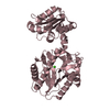
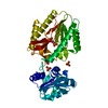
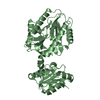
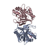
| Entry | Database: PDB / ID: 3n9w | ||||||
|---|---|---|---|---|---|---|---|
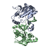
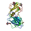
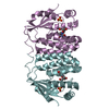
| Title | Crystal structure of 2-C-methyl-D-erythritol 4-phosphate cytidylyltransferase (IspD) in complex with 1,2-Propanediol | ||||||
 Components Components | 2-C-methyl-D-erythritol 4-phosphate cytidylyltransferase | ||||||
 Keywords Keywords | TRANSFERASE / YGBP / CYTIDYLYLTRANSFERASE / DEOXYXYLULOSE-5-PHOSPHATE PATHWAY (DXP) / ISOPRENOID BIOSYNTHESIS / 1 / 2-Propanediol / FBLD | ||||||
| Function / homology |  Function and homology information Function and homology information2-C-methyl-D-erythritol 4-phosphate cytidylyltransferase / 2-C-methyl-D-erythritol 4-phosphate cytidylyltransferase activity / isopentenyl diphosphate biosynthetic process, methylerythritol 4-phosphate pathway / magnesium ion binding / identical protein binding / cytosol Similarity search - Function | ||||||
| Biological species |  | ||||||
| Method |  X-RAY DIFFRACTION / SYNCHROTRON / MOLECULAR REPLACEMENT / Resolution: 1.9 Å X-RAY DIFFRACTION / SYNCHROTRON / MOLECULAR REPLACEMENT / Resolution: 1.9 Å | ||||||
 Authors Authors | Behnen, J. / Heine, A. / Klebe, G. | ||||||
 Citation Citation | Journal: Chemmedchem / Year: 2012 Title: Experimental and computational active site mapping as a starting point to fragment-based lead discovery. Authors: Behnen, J. / Koster, H. / Neudert, G. / Craan, T. / Heine, A. / Klebe, G. | ||||||
| History |
|
- Structure visualization
| Structure viewer | Molecule: MolmilJmol/JSmol |
|---|
- Downloads & links
-Download
| PDBx/mmCIF format | 3n9w.cif.gz | 92 KB | Display | PDBx/mmCIF format |
|---|---|---|---|---|
| PDB format | pdb3n9w.ent.gz | 69 KB | Display | PDB format |
| PDBx/mmJSON format | 3n9w.json.gz | Tree view | PDBx/mmJSON format | |
| Others |  Other downloads Other downloads |
-Validation report
| Arichive directory | https://data.pdbj.org/pub/pdb/validation_reports/n9/3n9wftp://data.pdbj.org/pub/pdb/validation_reports/n9/3n9w | HTTPS FTP |
|---|
-Related structure data
| Related structure data |  3ms3C 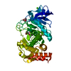 3msaC  3msfC 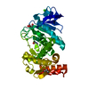 3msnC  3n21C 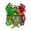 3n4aC  3nn7C 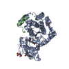 3nx8C 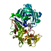 3pczC 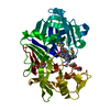 3prsC  3pvkC 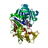 3pwwC  1vgtS S: Starting model for refinement C: citing same article ( |
|---|---|
| Similar structure data |


-Links
 PDBj
PDBj

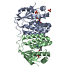
- Assembly
| Deposited unit | 
| ||||||||
|---|---|---|---|---|---|---|---|---|---|
| 1 |
| ||||||||
| Unit cell |
|
-Components
| #1: Protein | Mass: 25641.068 Da / Num. of mol.: 2 / Source method: isolated from a natural source / Source: (natural) References: UniProt: Q46893, 2-C-methyl-D-erythritol 4-phosphate cytidylyltransferase #2: Chemical |   Mass: 76.094 Da / Num. of mol.: 2 / Source method: obtained synthetically / Formula: C3H8O2 Mass: 76.094 Da / Num. of mol.: 2 / Source method: obtained synthetically / Formula: C3H8O2#3: Chemical | ChemComp-PGR / | 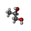  Mass: 76.094 Da / Num. of mol.: 1 / Source method: obtained synthetically / Formula: C3H8O2 Mass: 76.094 Da / Num. of mol.: 1 / Source method: obtained synthetically / Formula: C3H8O2#4: Water | ChemComp-HOH / |  Mass: 18.015 Da / Num. of mol.: 105 / Source method: isolated from a natural source / Formula: H2O Mass: 18.015 Da / Num. of mol.: 105 / Source method: isolated from a natural source / Formula: H2O |
|---|
-Experimental details
-Experiment
| Experiment | Method: X-RAY DIFFRACTION / Number of used crystals: 1 |
|---|
- Sample preparation
| Crystal | Density Matthews: 2.18 Å3/Da / Density % sol: 43.62 % |
|---|---|
| Crystal grow | Temperature: 277 K / Method: vapor diffusion, sitting drop / pH: 7.5 Details: 30% 1,2-propanediol, 20% PEG400, 0.1M HEPES, pH 7.5, VAPOR DIFFUSION, SITTING DROP, temperature 277K |
-Data collection
| Diffraction | Mean temperature: 110 K |
|---|---|
| Diffraction source | Source: SYNCHROTRON / Site: SLS  / Beamline: X06DA / Wavelength: 1 Å / Beamline: X06DA / Wavelength: 1 Å |
| Detector | Type: MARMOSAIC 225 mm CCD / Detector: CCD / Date: Aug 1, 2009 |
| Radiation | Monochromator: Bartels monochromator / Protocol: SINGLE WAVELENGTH / Monochromatic (M) / Laue (L): M / Scattering type: x-ray |
| Radiation wavelength | Wavelength: 1 Å / Relative weight: 1 |
| Reflection | Resolution: 1.9→25 Å / Num. all: 32991 / Num. obs: 32991 / % possible obs: 94.5 % / Observed criterion σ(F): 0 / Observed criterion σ(I): 0 / Redundancy: 3.3 % / Rsym value: 0.049 / Net I/σ(I): 21.8 |
| Reflection shell | Resolution: 1.9→1.93 Å / Redundancy: 2.9 % / Mean I/σ(I) obs: 2.9 / Rsym value: 0.287 / % possible all: 78 |
- Processing
| Software |
| |||||||||||||||||||||||||||||||||
|---|---|---|---|---|---|---|---|---|---|---|---|---|---|---|---|---|---|---|---|---|---|---|---|---|---|---|---|---|---|---|---|---|---|---|
| Refinement | Method to determine structure: MOLECULAR REPLACEMENT Starting model: PDB entry 1VGT Resolution: 1.9→10 Å / Num. parameters: 12823 / Num. restraintsaints: 16547 / Cross valid method: FREE R / σ(F): 0 / Stereochemistry target values: ENGH AND HUBER Details: ANISOTROPIC SCALING APPLIED BY THE METHOD OF PARKIN, MOEZZI & HOPE, J.APPL.CRYST.28(1995)53-56
| |||||||||||||||||||||||||||||||||
| Refine analyze | Num. disordered residues: 3 / Occupancy sum hydrogen: 3086 / Occupancy sum non hydrogen: 3177 | |||||||||||||||||||||||||||||||||
| Refinement step | Cycle: LAST / Resolution: 1.9→10 Å
| |||||||||||||||||||||||||||||||||
| Refine LS restraints |
| |||||||||||||||||||||||||||||||||
| LS refinement shell | Resolution: 1.9→1.93 Å / Num. reflection Rfree: 3287 |
