 Movie
Movie Controller

[English] 日本語
 Yorodumi
- PDB-2zxv: Crystal structure of putative acetyltransferase from T. thermophi... -
+ Open data

- Basic information
| Entry | Database: PDB / ID: 2zxv | ||||||
|---|---|---|---|---|---|---|---|
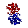
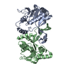
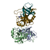
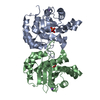
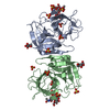
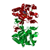
| Title | Crystal structure of putative acetyltransferase from T. thermophilus HB8 | ||||||
 Components Components | Putative uncharacterized protein TTHA1799 | ||||||
 Keywords Keywords | TRANSFERASE / alpha/beta protein / IY-substitution / Structural Genomics / NPPSFA / National Project on Protein Structural and Functional Analyses / RIKEN Structural Genomics/Proteomics Initiative / RSGI | ||||||
| Function / homology |  Function and homology information Function and homology informationacyltransferase activity, transferring groups other than amino-acyl groups / identical protein binding Similarity search - Function | ||||||
| Biological species |   Thermus thermophilus (bacteria) Thermus thermophilus (bacteria) | ||||||
| Method |  X-RAY DIFFRACTION / MOLECULAR REPLACEMENT / Resolution: 2.3 Å X-RAY DIFFRACTION / MOLECULAR REPLACEMENT / Resolution: 2.3 Å | ||||||
 Authors Authors | Murayama, K. / Kato-Murayama, M. / Terada, T. / Kuramitsu, S. / Shirouzu, M. / Yokoyama, S. / RIKEN Structural Genomics/Proteomics Initiative (RSGI) | ||||||
 Citation Citation | Journal: Structure / Year: 2009 Title: Genetic Encoding of 3-Iodo-l-Tyrosine in Escherichia coli for Single-Wavelength Anomalous Dispersion Phasing in Protein Crystallography Authors: Sakamoto, K. / Murayama, K. / Oki, K. / Iraha, F. / Kato-Murayama, M. / Takahashi, M. / Ohtake, K. / Kobayashi, T. / Kuramitsu, S. / Shirouzu, M. / Yokoyama, S. | ||||||
| History |
|
- Structure visualization
| Structure viewer | Molecule: MolmilJmol/JSmol |
|---|
- Downloads & links
-Download
| PDBx/mmCIF format | 2zxv.cif.gz | 161.9 KB | Display | PDBx/mmCIF format |
|---|---|---|---|---|
| PDB format | pdb2zxv.ent.gz | 131.4 KB | Display | PDB format |
| PDBx/mmJSON format | 2zxv.json.gz | Tree view | PDBx/mmJSON format | |
| Others |  Other downloads Other downloads |
-Validation report
| Arichive directory | https://data.pdbj.org/pub/pdb/validation_reports/zx/2zxvftp://data.pdbj.org/pub/pdb/validation_reports/zx/2zxv | HTTPS FTP |
|---|
-Related structure data
| Related structure data | 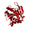 2z0zC 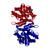 2z10SC C: citing same article ( S: Starting model for refinement |
|---|---|
| Similar structure data | |
| Other databases |

-Links
 PDBj
PDBj

- Assembly
| Deposited unit | 
| ||||||||
|---|---|---|---|---|---|---|---|---|---|
| 1 | 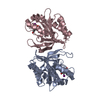
| ||||||||
| 2 | 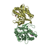
| ||||||||
| 3 | 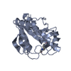
| ||||||||
| 4 | 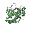
| ||||||||
| 5 | 
| ||||||||
| 6 | 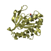
| ||||||||
| Unit cell |
|
-Components
| #1: Protein | Mass: 22288.467 Da / Num. of mol.: 4 / Mutation: Y35(IYR) Source method: isolated from a genetically manipulated source Source: (gene. exp.) Thermus thermophilus (bacteria) / Strain: HB8 / Gene: TTHA1799 / Plasmid: pET11a / Production host: References: UniProt: Q5SHD1, ribosomal-protein-alanine N-acetyltransferase #2: Water | ChemComp-HOH / |  Mass: 18.015 Da / Num. of mol.: 176 / Source method: isolated from a natural source / Formula: H2O Mass: 18.015 Da / Num. of mol.: 176 / Source method: isolated from a natural source / Formula: H2OHas protein modification | Y | |
|---|
-Experimental details
-Experiment
| Experiment | Method: X-RAY DIFFRACTION / Number of used crystals: 1 |
|---|
- Sample preparation
| Crystal | Density Matthews: 2.8 Å3/Da / Density % sol: 56.01 % |
|---|---|
| Crystal grow | Temperature: 293 K / Method: vapor diffusion, sitting drop / pH: 6.5 Details: 25% PEG 3350, 0.2M MgCl, 0.1M BisTris, pH 6.5, VAPOR DIFFUSION, SITTING DROP, temperature 293K |
-Data collection
| Diffraction | Mean temperature: 100 K |
|---|---|
| Diffraction source | Source: ROTATING ANODE / Type: RIGAKU FR-E+ SUPERBRIGHT / Wavelength: 1.5418 Å |
| Detector | Type: RIGAKU RAXIS IV++ / Detector: IMAGE PLATE / Date: Apr 12, 2005 |
| Radiation | Protocol: SINGLE WAVELENGTH / Monochromatic (M) / Laue (L): M / Scattering type: x-ray |
| Radiation wavelength | Wavelength: 1.5418 Å / Relative weight: 1 |
| Reflection | Resolution: 2.3→50 Å / Num. obs: 42723 / % possible obs: 97.8 % / Observed criterion σ(I): -3 / Redundancy: 14.5 % / Biso Wilson estimate: 23.3 Å2 / Rsym value: 0.089 / Net I/σ(I): 28.7 |
| Reflection shell | Resolution: 2.3→2.38 Å / Redundancy: 10.7 % / Mean I/σ(I) obs: 8.5 / Rsym value: 0.296 / % possible all: 82.3 |
- Processing
| Software |
| ||||||||||||||||||||||||||||||||||||
|---|---|---|---|---|---|---|---|---|---|---|---|---|---|---|---|---|---|---|---|---|---|---|---|---|---|---|---|---|---|---|---|---|---|---|---|---|---|
| Refinement | Method to determine structure: MOLECULAR REPLACEMENT Starting model: Pdb entry 2Z10 Resolution: 2.3→30.13 Å / Rfactor Rfree error: 0.006 / Data cutoff high absF: 2686186.95 / Data cutoff low absF: 0 / Isotropic thermal model: RESTRAINED / Cross valid method: THROUGHOUT / σ(F): 0 / Stereochemistry target values: Engh & Huber Details: THIS IS A TWINNED DATA. THE TWINNING OPERATOR IS (H,K,L) -> (h, -k, -l) AND THE TWINNING FRACTION is 0.443
| ||||||||||||||||||||||||||||||||||||
| Solvent computation | Solvent model: FLAT MODEL / Bsol: 28.3764 Å2 / ksol: 0.323298 e/Å3 | ||||||||||||||||||||||||||||||||||||
| Displacement parameters | Biso mean: 39.4 Å2
| ||||||||||||||||||||||||||||||||||||
| Refine analyze |
| ||||||||||||||||||||||||||||||||||||
| Refinement step | Cycle: LAST / Resolution: 2.3→30.13 Å
| ||||||||||||||||||||||||||||||||||||
| Refine LS restraints |
| ||||||||||||||||||||||||||||||||||||
| LS refinement shell | Resolution: 2.3→2.44 Å / Rfactor Rfree error: 0.018 / Total num. of bins used: 6
| ||||||||||||||||||||||||||||||||||||
| Xplor file |
|
