 Movie
Movie Controller


+ Open data

- Basic information
| Entry | Database: PDB / ID: 3pvk | |||||||||
|---|---|---|---|---|---|---|---|---|---|---|
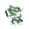
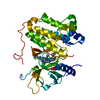
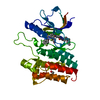
| Title | Secreted aspartic protease 2 in complex with benzamidine | |||||||||
 Components Components | Candidapepsin-2 | |||||||||
 Keywords Keywords | HYDROLASE | |||||||||
| Function / homology |  Function and homology information Function and homology informationprotein catabolic process => GO:0030163 / : / : / protein metabolic process / candidapepsin / : / : / fungal-type cell wall organization / adhesion of symbiont to host / fungal-type cell wall ...protein catabolic process => GO:0030163 / : / : / protein metabolic process / candidapepsin / : / : / fungal-type cell wall organization / adhesion of symbiont to host / fungal-type cell wall / protein catabolic process / extracellular vesicle / aspartic-type endopeptidase activity / proteolysis / extracellular region / metal ion binding Similarity search - Function | |||||||||
| Biological species |  Candida albicans (yeast) Candida albicans (yeast) | |||||||||
| Method |  X-RAY DIFFRACTION / SYNCHROTRON / MOLECULAR REPLACEMENT / Resolution: 1.27 Å X-RAY DIFFRACTION / SYNCHROTRON / MOLECULAR REPLACEMENT / Resolution: 1.27 Å | |||||||||
 Authors Authors | Koester, H. / Heine, A. / Klebe, G. | |||||||||
 Citation Citation | Journal: Chemmedchem / Year: 2012 Title: Experimental and computational active site mapping as a starting point to fragment-based lead discovery. Authors: Behnen, J. / Koster, H. / Neudert, G. / Craan, T. / Heine, A. / Klebe, G. | |||||||||
| History |
|
- Structure visualization
| Structure viewer | Molecule: MolmilJmol/JSmol |
|---|
- Downloads & links
-Download
| PDBx/mmCIF format | 3pvk.cif.gz | 85 KB | Display | PDBx/mmCIF format |
|---|---|---|---|---|
| PDB format | pdb3pvk.ent.gz | 61.8 KB | Display | PDB format |
| PDBx/mmJSON format | 3pvk.json.gz | Tree view | PDBx/mmJSON format | |
| Others |  Other downloads Other downloads |
-Validation report
| Arichive directory | https://data.pdbj.org/pub/pdb/validation_reports/pv/3pvkftp://data.pdbj.org/pub/pdb/validation_reports/pv/3pvk | HTTPS FTP |
|---|
-Related structure data
| Related structure data |  3ms3C 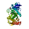 3msaC  3msfC 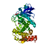 3msnC  3n21C 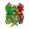 3n4aC 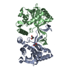 3n9wC  3nn7C 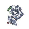 3nx8C 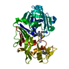 3pczC 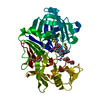 3prsC 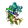 3pwwC 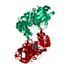 1eagS S: Starting model for refinement C: citing same article ( |
|---|---|
| Similar structure data |


-Links
 PDBj
PDBj


- Assembly
| Deposited unit | 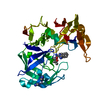
| ||||||||
|---|---|---|---|---|---|---|---|---|---|
| 1 |
| ||||||||
| Unit cell |
|
-Components
-Protein , 1 types, 1 molecules A
| #1: Protein | Mass: 36357.723 Da / Num. of mol.: 1 / Fragment: UNP residues 57-398 / Source method: isolated from a natural source / Source: (natural) Candida albicans (yeast)References: UniProt: P28871, UniProt: P0CS83*PLUS, candidapepsin |
|---|
-Non-polymers , 5 types, 338 molecules 


| #2: Chemical | ChemComp-ZN /  Mass: 65.409 Da / Num. of mol.: 1 / Source method: obtained synthetically / Formula: Zn Mass: 65.409 Da / Num. of mol.: 1 / Source method: obtained synthetically / Formula: Zn | ||||||
|---|---|---|---|---|---|---|---|
| #3: Chemical |  Mass: 120.152 Da / Num. of mol.: 2 / Source method: obtained synthetically / Formula: C7H8N2 Mass: 120.152 Da / Num. of mol.: 2 / Source method: obtained synthetically / Formula: C7H8N2#4: Chemical | ChemComp-MPD / ( |  Mass: 118.174 Da / Num. of mol.: 1 / Source method: obtained synthetically / Formula: C6H14O2 / Comment: precipitant*YM Mass: 118.174 Da / Num. of mol.: 1 / Source method: obtained synthetically / Formula: C6H14O2 / Comment: precipitant*YM#5: Chemical |  Mass: 69.085 Da / Num. of mol.: 2 / Source method: obtained synthetically / Formula: C3H5N2 Mass: 69.085 Da / Num. of mol.: 2 / Source method: obtained synthetically / Formula: C3H5N2#6: Water | ChemComp-HOH / | Mass: 18.015 Da / Num. of mol.: 332 / Source method: isolated from a natural source / Formula: H2O |
-Details
| Has protein modification | Y |
|---|
-Experimental details
-Experiment
| Experiment | Method: X-RAY DIFFRACTION / Number of used crystals: 1 |
|---|
- Sample preparation
| Crystal | Density Matthews: 2.12 Å3/Da / Density % sol: 42.1 % |
|---|---|
| Crystal grow | Temperature: 289 K / Method: vapor diffusion, sitting drop / pH: 6.5 Details: 9 mM ZnAc, 16% PEG 8000, 0.1M imidazole/malate buffer pH 6.5, VAPOR DIFFUSION, SITTING DROP, temperature 289K |
-Data collection
| Diffraction | Mean temperature: 100 K |
|---|---|
| Diffraction source | Source: SYNCHROTRON / Site: SLS  / Beamline: X06DA / Wavelength: 1 Å / Beamline: X06DA / Wavelength: 1 Å |
| Detector | Type: MARMOSAIC 225 mm CCD / Detector: CCD / Date: Jul 31, 2009 / Details: mirrors |
| Radiation | Monochromator: Bertels Monochromator with dual channel cut crystals (DCCM) in (+--+) geometry Protocol: SINGLE WAVELENGTH / Monochromatic (M) / Laue (L): M / Scattering type: x-ray |
| Radiation wavelength | Wavelength: 1 Å / Relative weight: 1 |
| Reflection | Resolution: 1.27→40 Å / Num. all: 79920 / Num. obs: 79920 / % possible obs: 100 % / Redundancy: 6.8 % / Rsym value: 0.071 / Net I/σ(I): 26.2 |
| Reflection shell | Resolution: 1.27→1.29 Å / Redundancy: 3.7 % / Mean I/σ(I) obs: 2.9 / Num. unique all: 3341 / Rsym value: 0.418 / % possible all: 100 |
- Processing
| Software |
| |||||||||||||||||||||||||||||||||||||||||||||||||||||||||||||||||||||||||||||
|---|---|---|---|---|---|---|---|---|---|---|---|---|---|---|---|---|---|---|---|---|---|---|---|---|---|---|---|---|---|---|---|---|---|---|---|---|---|---|---|---|---|---|---|---|---|---|---|---|---|---|---|---|---|---|---|---|---|---|---|---|---|---|---|---|---|---|---|---|---|---|---|---|---|---|---|---|---|---|
| Refinement | Method to determine structure: MOLECULAR REPLACEMENT Starting model: 1EAG Resolution: 1.27→38.999 Å / SU ML: 0.12 / σ(F): 0 / Stereochemistry target values: ML
| |||||||||||||||||||||||||||||||||||||||||||||||||||||||||||||||||||||||||||||
| Solvent computation | Shrinkage radii: 0.9 Å / VDW probe radii: 1.11 Å / Solvent model: FLAT BULK SOLVENT MODEL / Bsol: 39.22 Å2 / ksol: 0.358 e/Å3 | |||||||||||||||||||||||||||||||||||||||||||||||||||||||||||||||||||||||||||||
| Displacement parameters |
| |||||||||||||||||||||||||||||||||||||||||||||||||||||||||||||||||||||||||||||
| Refinement step | Cycle: LAST / Resolution: 1.27→38.999 Å
| |||||||||||||||||||||||||||||||||||||||||||||||||||||||||||||||||||||||||||||
| Refine LS restraints |
| |||||||||||||||||||||||||||||||||||||||||||||||||||||||||||||||||||||||||||||
| LS refinement shell |
|
