 Movie
Movie Controller

[English] 日本語
 Yorodumi
- PDB-5d7a: Crystal structure of the kinase domain of TRAF2 and NCK-interacti... -
+ Open data

- Basic information
| Entry | Database: PDB / ID: 5d7a | ||||||
|---|---|---|---|---|---|---|---|
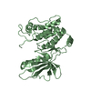
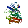
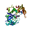
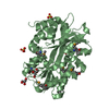
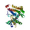
| Title | Crystal structure of the kinase domain of TRAF2 and NCK-interacting protein kinase with NCB-0846 | ||||||
 Components Components | TRAF2 and NCK-interacting protein kinase | ||||||
 Keywords Keywords | TRANSFERASE/TRANSFERASE INHIBITOR / Kinase / TRANSFERASE-TRANSFERASE INHIBITOR complex | ||||||
| Function / homology |  Function and homology information Function and homology informationpositive regulation of microvillus assembly / regulation of dendrite morphogenesis / regulation of MAPK cascade / postsynaptic density, intracellular component / neuron projection morphogenesis / cytoskeleton organization / recycling endosome / positive regulation of JNK cascade / Wnt signaling pathway / protein autophosphorylation ...positive regulation of microvillus assembly / regulation of dendrite morphogenesis / regulation of MAPK cascade / postsynaptic density, intracellular component / neuron projection morphogenesis / cytoskeleton organization / recycling endosome / positive regulation of JNK cascade / Wnt signaling pathway / protein autophosphorylation / MAPK cascade / presynapse / actin cytoskeleton organization / Oxidative Stress Induced Senescence / cytoskeleton / protein phosphorylation / protein kinase activity / non-specific serine/threonine protein kinase / apical plasma membrane / intracellular signal transduction / protein serine kinase activity / protein serine/threonine kinase activity / glutamatergic synapse / extracellular exosome / nucleoplasm / ATP binding / nucleus / cytoplasm / cytosol Similarity search - Function | ||||||
| Biological species |  Homo sapiens (human) Homo sapiens (human) | ||||||
| Method |  X-RAY DIFFRACTION / SYNCHROTRON / MOLECULAR REPLACEMENT / Resolution: 2.9 Å X-RAY DIFFRACTION / SYNCHROTRON / MOLECULAR REPLACEMENT / Resolution: 2.9 Å | ||||||
 Authors Authors | Ohbayashi, N. / Kukimoto-Niino, M. / Yamada, T. / Shirouzu, M. | ||||||
 Citation Citation | Journal: Nat Commun / Year: 2016 Title: TNIK inhibition abrogates colorectal cancer stemness Authors: Masuda, M. / Uno, Y. / Ohbayashi, N. / Ohata, H. / Mimata, A. / Kukimoto-Niino, M. / Moriyama, H. / Kashimoto, S. / Inoue, T. / Goto, N. / Okamoto, K. / Shirouzu, M. / Sawa, M. / Yamada, T. | ||||||
| History |
|
- Structure visualization
| Structure viewer | Molecule: MolmilJmol/JSmol |
|---|
- Downloads & links
-Download
| PDBx/mmCIF format | 5d7a.cif.gz | 183.4 KB | Display | PDBx/mmCIF format |
|---|---|---|---|---|
| PDB format | pdb5d7a.ent.gz | 146.2 KB | Display | PDB format |
| PDBx/mmJSON format | 5d7a.json.gz | Tree view | PDBx/mmJSON format | |
| Others |  Other downloads Other downloads |
-Validation report
| Arichive directory | https://data.pdbj.org/pub/pdb/validation_reports/d7/5d7aftp://data.pdbj.org/pub/pdb/validation_reports/d7/5d7a | HTTPS FTP |
|---|
-Related structure data
| Related structure data |  5ax9C  5cwzC  2x7fS C: citing same article ( S: Starting model for refinement |
|---|---|
| Similar structure data |


-Links
 PDBj
PDBj- Assembly
| Deposited unit | 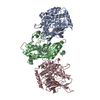
| ||||||||
|---|---|---|---|---|---|---|---|---|---|
| 1 | 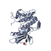
| ||||||||
| 2 | 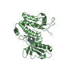
| ||||||||
| 3 | 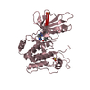
| ||||||||
| Unit cell |
|
-Components
| #1: Protein | Mass: 35076.328 Da / Num. of mol.: 3 Source method: isolated from a genetically manipulated source Source: (gene. exp.) Homo sapiens (human) / Gene: TNIK, KIAA0551 / Production host:  Baculovirus expression vector pFastBac1-HM Baculovirus expression vector pFastBac1-HMReferences: UniProt: Q9UKE5, non-specific serine/threonine protein kinase #2: Chemical |   Mass: 375.424 Da / Num. of mol.: 3 / Source method: obtained synthetically / Formula: C21H21N5O2 Mass: 375.424 Da / Num. of mol.: 3 / Source method: obtained synthetically / Formula: C21H21N5O2#3: Chemical |   Mass: 96.063 Da / Num. of mol.: 3 / Source method: obtained synthetically / Formula: SO4 Mass: 96.063 Da / Num. of mol.: 3 / Source method: obtained synthetically / Formula: SO4#4: Water | ChemComp-HOH / |  Mass: 18.015 Da / Num. of mol.: 7 / Source method: isolated from a natural source / Formula: H2O Mass: 18.015 Da / Num. of mol.: 7 / Source method: isolated from a natural source / Formula: H2O |
|---|
-Experimental details
-Experiment
| Experiment | Method: X-RAY DIFFRACTION |
|---|
- Sample preparation
| Crystal | Density Matthews: 3.13 Å3/Da / Density % sol: 60.71 % |
|---|---|
| Crystal grow | Temperature: 293 K / Method: vapor diffusion, sitting drop / pH: 5.6 Details: 0.2M Ammonium sulfate 0.1M Bis-tris (5.6) 15% PEG3350 7% Ethyleneglycol |
-Data collection
| Diffraction | Mean temperature: 100 K |
|---|---|
| Diffraction source | Source: SYNCHROTRON / Site: SPring-8  / Beamline: BL26B2 / Wavelength: 1 Å / Beamline: BL26B2 / Wavelength: 1 Å |
| Detector | Type: MARMOSAIC 225 mm CCD / Detector: CCD / Date: Apr 21, 2015 |
| Radiation | Protocol: SINGLE WAVELENGTH / Monochromatic (M) / Laue (L): M / Scattering type: x-ray |
| Radiation wavelength | Wavelength: 1 Å / Relative weight: 1 |
| Reflection | Resolution: 2.9→46.92 Å / Num. obs: 28803 / % possible obs: 100 % / Redundancy: 7.7 % / Rmerge(I) obs: 0.117 / Net I/σ(I): 15.8 |
| Reflection shell | Resolution: 2.9→3.08 Å / Redundancy: 7.8 % / Mean I/σ(I) obs: 2.7 / % possible all: 100 |
- Processing
| Software |
| |||||||||||||||||||||||||||||||||||||||||||||||||||||||||||||||||||||||||||||
|---|---|---|---|---|---|---|---|---|---|---|---|---|---|---|---|---|---|---|---|---|---|---|---|---|---|---|---|---|---|---|---|---|---|---|---|---|---|---|---|---|---|---|---|---|---|---|---|---|---|---|---|---|---|---|---|---|---|---|---|---|---|---|---|---|---|---|---|---|---|---|---|---|---|---|---|---|---|---|
| Refinement | Method to determine structure: MOLECULAR REPLACEMENT Starting model: 2X7F Resolution: 2.9→46.92 Å / SU ML: 0.41 / Cross valid method: FREE R-VALUE / σ(F): 1.35 / Phase error: 26.02 / Stereochemistry target values: ML
| |||||||||||||||||||||||||||||||||||||||||||||||||||||||||||||||||||||||||||||
| Solvent computation | Shrinkage radii: 0.9 Å / VDW probe radii: 1.11 Å / Solvent model: FLAT BULK SOLVENT MODEL | |||||||||||||||||||||||||||||||||||||||||||||||||||||||||||||||||||||||||||||
| Refinement step | Cycle: LAST / Resolution: 2.9→46.92 Å
| |||||||||||||||||||||||||||||||||||||||||||||||||||||||||||||||||||||||||||||
| Refine LS restraints |
| |||||||||||||||||||||||||||||||||||||||||||||||||||||||||||||||||||||||||||||
| LS refinement shell |
|
