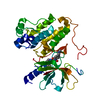
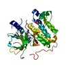
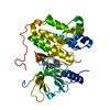
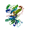
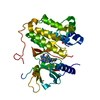
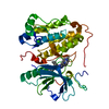
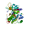
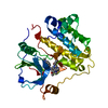
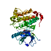
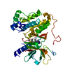
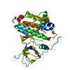
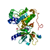
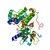
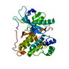
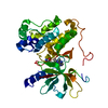
Entry Database : PDB / ID : 5hczTitle EGFR kinase domain mutant "TMLR" with 3-azetidinyl azaindazole compound 21 Epidermal growth factor receptor Keywords / / / Function / homology Function Domain/homology Component
/ / / / / / / / / / / / / / / / / / / / / / / / / / / / / / / / / / / / / / / / / / / / / / / / / / / / / / / / / / / / / / / / / / / / / / / / / / / / / / / / / / / / / / / / / / / / / / / / / / / / / / / / / / / / / / / / / / / / / / / / / / / / / / / / / / / / / / / / / / / / / / / / / / / / / Biological species Homo sapiens (human)Method / / / Resolution : 2.62 Å Authors Eigenbrot, C. / Yu, C. Journal : J.Med.Chem. / Year : 2016Title : Discovery of a Noncovalent, Mutant-Selective Epidermal Growth Factor Receptor Inhibitor.Authors: Chan, B.K. / Hanan, E.J. / Bowman, K.K. / Bryan, M.C. / Burdick, D. / Chan, E. / Chen, Y. / Clausen, S. / Dela Vega, T. / Dotson, J. / Eigenbrot, C. / Elliott, R.L. / Heald, R.A. / Jackson, ... Authors : Chan, B.K. / Hanan, E.J. / Bowman, K.K. / Bryan, M.C. / Burdick, D. / Chan, E. / Chen, Y. / Clausen, S. / Dela Vega, T. / Dotson, J. / Eigenbrot, C. / Elliott, R.L. / Heald, R.A. / Jackson, P.S. / Knight, J.D. / La, H. / Lainchbury, M.D. / Malek, S. / Purkey, H.E. / Schaefer, G. / Schmidt, S. / Seward, E.M. / Sideris, S. / Shao, L. / Wang, S. / Yeap, S.K. / Yen, I. / Yu, C. / Heffron, T.P. History Deposition Jan 4, 2016 Deposition site / Processing site Revision 1.0 Sep 7, 2016 Provider / Type Revision 1.1 Oct 26, 2016 Group Revision 1.2 Sep 27, 2023 Group Data collection / Database references ... Data collection / Database references / Derived calculations / Refinement description Category chem_comp_atom / chem_comp_bond ... chem_comp_atom / chem_comp_bond / citation / database_2 / pdbx_initial_refinement_model / pdbx_struct_oper_list Item _citation.journal_id_CSD / _database_2.pdbx_DOI ... _citation.journal_id_CSD / _database_2.pdbx_DOI / _database_2.pdbx_database_accession / _pdbx_struct_oper_list.symmetry_operation
Show all Show less
 Movie
Movie Controller

 Yorodumi
 Open data
 Basic information
 Components
 Keywords
 Function and homology information
 Homo sapiens (human)
 X-RAY DIFFRACTION /
  Authors
 Citation
 Structure visualization
 Downloads & links
 Other downloads
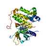


 PDBj
PDBj


 Assembly

 Spodoptera frugiperda (fall armyworm)
 Mass: 96.063 Da / Num. of mol.: 1 / Source method: obtained synthetically / Formula: SO4
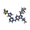
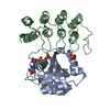
 Mass: 551.664 Da / Num. of mol.: 1 / Source method: obtained synthetically / Formula: C26H33N9O3S
 Mass: 18.015 Da / Num. of mol.: 10 / Source method: isolated from a natural source / Formula: H2O
 Sample preparation
 / Beamline: BL12-2 / Wavelength: 0.9795 Å
 Processing