histone H2AK127 ubiquitin ligase activity / histone H2AK129 ubiquitin ligase activity / Defective DNA double strand break response due to BRCA1 loss of function / Defective DNA double strand break response due to BARD1 loss of function / BRCA1-BARD1 complex / BRCA1-C complex / BRCA1-B complex / BRCA1-A complex / negative regulation of centriole replication / nuclear ubiquitin ligase complex ...histone H2AK127 ubiquitin ligase activity / histone H2AK129 ubiquitin ligase activity / Defective DNA double strand break response due to BRCA1 loss of function / Defective DNA double strand break response due to BARD1 loss of function / BRCA1-BARD1 complex / BRCA1-C complex / BRCA1-B complex / BRCA1-A complex / negative regulation of centriole replication / nuclear ubiquitin ligase complex / random inactivation of X chromosome / chordate embryonic development / gamma-tubulin ring complex / negative regulation of intracellular estrogen receptor signaling pathway / ubiquitin-modified histone reader activity / cellular response to indole-3-methanol / DNA strand resection involved in replication fork processing / homologous recombination / negative regulation of fatty acid biosynthetic process / Regulation of MITF-M-dependent genes involved in DNA replication, damage repair and senescence / lateral element / mitotic G2/M transition checkpoint / XY body / Impaired BRCA2 binding to PALB2 / regulation of DNA damage checkpoint / DNA repair complex / protein K6-linked ubiquitination / RNA polymerase binding / DNA damage tolerance / centrosome cycle / negative regulation of gene expression via chromosomal CpG island methylation / HDR through Single Strand Annealing (SSA) / response to ionizing radiation / Homologous DNA Pairing and Strand Exchange / Defective homologous recombination repair (HRR) due to BRCA1 loss of function / Defective HDR through Homologous Recombination Repair (HRR) due to PALB2 loss of BRCA1 binding function / Defective HDR through Homologous Recombination Repair (HRR) due to PALB2 loss of BRCA2/RAD51/RAD51C binding function / Resolution of D-loop Structures through Synthesis-Dependent Strand Annealing (SDSA) / mitotic G2 DNA damage checkpoint signaling / Resolution of D-loop Structures through Holliday Junction Intermediates / Transcriptional Regulation by E2F6 / negative regulation of cell cycle / Impaired BRCA2 binding to RAD51 / negative regulation of reactive oxygen species metabolic process / positive regulation of vascular endothelial growth factor production / Presynaptic phase of homologous DNA pairing and strand exchange / ubiquitin ligase complex / regulation of DNA repair / negative regulation of extrinsic apoptotic signaling pathway via death domain receptors / SUMOylation of DNA damage response and repair proteins / protein autoubiquitination / male germ cell nucleus / Meiotic synapsis / positive regulation of DNA repair / cellular response to ionizing radiation / TP53 Regulates Transcription of DNA Repair Genes / chromosome segregation / Nonhomologous End-Joining (NHEJ) / tubulin binding / negative regulation of cell growth / RING-type E3 ubiquitin transferase / G2/M DNA damage checkpoint / double-strand break repair via homologous recombination / intrinsic apoptotic signaling pathway in response to DNA damage / cellular response to tumor necrosis factor / Meiotic recombination / HDR through Homologous Recombination (HRR) / fatty acid biosynthetic process / Metalloprotease DUBs / positive regulation of angiogenesis / p53 binding / ubiquitin-protein transferase activity / KEAP1-NFE2L2 pathway / double-strand break repair / chromosome / Recruitment and ATM-mediated phosphorylation of repair and signaling proteins at DNA double strand breaks / Neddylation / Processing of DNA double-strand break ends / Regulation of TP53 Activity through Phosphorylation / damaged DNA binding / transcription coactivator activity / regulation of cell cycle / transcription cis-regulatory region binding / nuclear body / protein ubiquitination / chromatin remodeling / ribonucleoprotein complex / negative regulation of DNA-templated transcription / DNA repair / positive regulation of gene expression / ubiquitin protein ligase binding / regulation of transcription by RNA polymerase II / DNA damage response / positive regulation of DNA-templated transcription / enzyme binding / positive regulation of transcription by RNA polymerase II / protein-containing complex / DNA binding / RNA binding / zinc ion binding Similarity search - Function
Breast cancer type 1 susceptibility protein (BRCA1) / BRCA1, serine-rich domain / BRCA1-associated / Serine-rich domain associated with BRCT / BRCT domain / Zinc finger, C3HC4 RING-type / Zinc finger, C3HC4 type (RING finger) / BRCA1 C Terminus (BRCT) domain / breast cancer carboxy-terminal domain / BRCT domain profile. ...Breast cancer type 1 susceptibility protein (BRCA1) / BRCA1, serine-rich domain / BRCA1-associated / Serine-rich domain associated with BRCT / BRCT domain / Zinc finger, C3HC4 RING-type / Zinc finger, C3HC4 type (RING finger) / BRCA1 C Terminus (BRCT) domain / breast cancer carboxy-terminal domain / BRCT domain profile. / BRCT domain / BRCT domain superfamily / Zinc finger, RING-type, conserved site / Zinc finger RING-type signature. / Ring finger / Zinc finger RING-type profile. / Zinc finger, RING-type / Zinc finger, RING/FYVE/PHD-type / Rossmann fold / 3-Layer(aba) Sandwich / Alpha Beta Similarity search - Domain/homology
Breastcancertype1susceptibilityprotein / RING finger protein 53
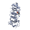
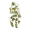
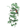
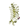
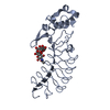
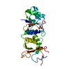
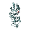
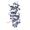
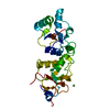
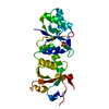
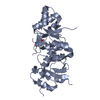
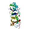
Mass: 24662.434 Da / Num. of mol.: 1 / Fragment: BRCT Domain (UNP residues 1646 to 1859) Source method: isolated from a genetically manipulated source Source: (gene. exp.) Homo sapiens (human) / Gene: BRCA1, RNF53 / Plasmid: pLM1-CD6 / Production host: Escherichia coli (E. coli) / Strain (production host): BL21 Gold / References: UniProt: P38398
#2: Protein/peptide
phosphopeptide
Mass: 528.472 Da / Num. of mol.: 1 / Source method: obtained synthetically Details: This sequence occurs naturally in humans in the BACH1/BRIP1 protein. The peptide is chemically synthesized.
In the structure databanks used in Yorodumi, some data are registered as the other names, "COVID-19 virus" and "2019-nCoV". Here are the details of the virus and the list of structure data.
Jan 31, 2019. EMDB accession codes are about to change! (news from PDBe EMDB page)
EMDB accession codes are about to change! (news from PDBe EMDB page)
The allocation of 4 digits for EMDB accession codes will soon come to an end. Whilst these codes will remain in use, new EMDB accession codes will include an additional digit and will expand incrementally as the available range of codes is exhausted. The current 4-digit format prefixed with “EMD-” (i.e. EMD-XXXX) will advance to a 5-digit format (i.e. EMD-XXXXX), and so on. It is currently estimated that the 4-digit codes will be depleted around Spring 2019, at which point the 5-digit format will come into force.
The EM Navigator/Yorodumi systems omit the EMD- prefix.
Related info.:Q: What is EMD? / ID/Accession-code notation in Yorodumi/EM Navigator
Yorodumi is a browser for structure data from EMDB, PDB, SASBDB, etc.
This page is also the successor to EM Navigator detail page, and also detail information page/front-end page for Omokage search.
The word "yorodu" (or yorozu) is an old Japanese word meaning "ten thousand". "mi" (miru) is to see.
Related info.:EMDB / PDB / SASBDB / Comparison of 3 databanks / Yorodumi Search / Aug 31, 2016. New EM Navigator & Yorodumi / Yorodumi Papers / Jmol/JSmol / Function and homology information / Changes in new EM Navigator and Yorodumi
 Movie
Movie Controller

 Yorodumi
 Open data
 Basic information
 Components
 Keywords
 Function and homology information
 Homo sapiens (human)
 X-RAY DIFFRACTION /
  Authors
 Citation
 Structure visualization
 Downloads & links
 Other downloads
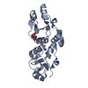


 PDBj
PDBj


 Assembly


 Mass: 58.693 Da / Num. of mol.: 1 / Source method: obtained synthetically / Formula: Ni
 Mass: 35.453 Da / Num. of mol.: 1 / Source method: obtained synthetically / Formula: Cl
 Mass: 18.015 Da / Num. of mol.: 18 / Source method: isolated from a natural source / Formula: H2O
 Sample preparation
 / Beamline: 08ID-1 / Wavelength: 0.97934 Å
 Processing