[English] 日本語
Yorodumi
- PDB-3k05: The crystal structure of MDC1 BRCT T2067D in complex with a minim... -

+
Open data


ID or keywords:

Loading...

-
Basic information

Entry
Database: PDB / ID: 3k05
Title
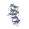
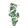
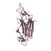
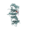
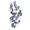
The crystal structure of MDC1 BRCT T2067D in complex with a minimal recognition tetrapeptide with an amidated C-terminus
Components
  • Mediator of DNA damage checkpoint protein 1
  • phospho peptide
KeywordsPROTEIN BINDING / BRCT Domain / protein-peptide complex / phospho protein binding / H2AX / DNA damage response / Cell cycle / DNA damage / DNA repair / Nucleus / Phosphoprotein
Function / homology
Function and homology information


chromatin-protein adaptor activity / DNA replication checkpoint signaling / protein localization to site of double-strand break / mitotic intra-S DNA damage checkpoint signaling / positive regulation of transcription initiation by RNA polymerase II / SUMOylation of DNA damage response and repair proteins / histone reader activity / TP53 Regulates Transcription of DNA Repair Genes / Nonhomologous End-Joining (NHEJ) / G2/M DNA damage checkpoint ...chromatin-protein adaptor activity / DNA replication checkpoint signaling / protein localization to site of double-strand break / mitotic intra-S DNA damage checkpoint signaling / positive regulation of transcription initiation by RNA polymerase II / SUMOylation of DNA damage response and repair proteins / histone reader activity / TP53 Regulates Transcription of DNA Repair Genes / Nonhomologous End-Joining (NHEJ) / G2/M DNA damage checkpoint / Recruitment and ATM-mediated phosphorylation of repair and signaling proteins at DNA double strand breaks / chromosome / site of double-strand break / Processing of DNA double-strand break ends / nuclear body / DNA repair / focal adhesion / DNA damage response / nucleoplasm / nucleus
Similarity search - Function
Regulator of Ty1 transposition protein 107 BRCT domain / BRCT domain / BRCT domain, a BRCA1 C-terminus domain / Forkhead associated domain / Forkhead-associated (FHA) domain profile. / FHA domain / Forkhead-associated (FHA) domain / SMAD/FHA domain superfamily / BRCT domain profile. / BRCT domain ...Regulator of Ty1 transposition protein 107 BRCT domain / BRCT domain / BRCT domain, a BRCA1 C-terminus domain / Forkhead associated domain / Forkhead-associated (FHA) domain profile. / FHA domain / Forkhead-associated (FHA) domain / SMAD/FHA domain superfamily / BRCT domain profile. / BRCT domain / BRCT domain superfamily / Rossmann fold / 3-Layer(aba) Sandwich / Alpha Beta
Similarity search - Domain/homology
Mediator of DNA damage checkpoint protein 1
Similarity search - Component
Biological speciesHomo sapiens (human)
MethodX-RAY DIFFRACTION / SYNCHROTRON / MOLECULAR REPLACEMENT / molecular replacement / Resolution: 1.33 Å
AuthorsCampbell, S.J. / Edwards, R.A. / Glover, J.N.
CitationJournal: Structure / Year: 2010
Title: Comparison of the Structures and Peptide Binding Specificities of the BRCT Domains of MDC1 and BRCA1
Authors: Campbell, S.J. / Edwards, R.A. / Glover, J.N.
History
DepositionSep 24, 2009Deposition site: RCSB / Processing site: RCSB
Revision 1.0Mar 2, 2010Provider: repository / Type: Initial release
Revision 1.1Jul 13, 2011Group: Version format compliance
Revision 1.2Nov 1, 2017Group: Refinement description / Category: software
Revision 1.3Oct 13, 2021Group: Database references / Derived calculations
Category: database_2 / struct_conn ...database_2 / struct_conn / struct_ref_seq / struct_ref_seq_dif / struct_site
Item: _database_2.pdbx_DOI / _database_2.pdbx_database_accession ..._database_2.pdbx_DOI / _database_2.pdbx_database_accession / _struct_conn.pdbx_leaving_atom_flag / _struct_ref_seq.db_align_beg / _struct_ref_seq.db_align_end / _struct_ref_seq_dif.details / _struct_site.pdbx_auth_asym_id / _struct_site.pdbx_auth_comp_id / _struct_site.pdbx_auth_seq_id

-
Structure visualization

Structure viewerMolecule:
MolmilJmol/JSmol

Downloads & links

-
Assembly

Deposited unit
A: Mediator of DNA damage checkpoint protein 1
B: Mediator of DNA damage checkpoint protein 1
C: phospho peptide
D: phospho peptide
hetero molecules


Theoretical massNumber of molelcules
Total (without water)45,2888
Polymers44,9204
Non-polymers3684
Water8,431468
1
A: Mediator of DNA damage checkpoint protein 1
C: phospho peptide
hetero molecules


Theoretical massNumber of molelcules
Total (without water)22,5523
Polymers22,4602
Non-polymers921
Water362
TypeNameSymmetry operationNumber
identity operation1_555x,y,z1
Buried area860 Å2
ΔGint-8 kcal/mol
Surface area9780 Å2
MethodPISA
2
B: Mediator of DNA damage checkpoint protein 1
D: phospho peptide
hetero molecules


Theoretical massNumber of molelcules
Total (without water)22,7365
Polymers22,4602
Non-polymers2763
Water362
TypeNameSymmetry operationNumber
identity operation1_555x,y,z1
Buried area1380 Å2
ΔGint-8 kcal/mol
Surface area9950 Å2
MethodPISA
Unit cell
Length a, b, c (Å)55.642, 74.580, 103.408
Angle α, β, γ (deg.)90.00, 90.00, 90.00
Int Tables number19
Space group name H-MP212121
DetailsThe biological unit is a monomer bound to a phospho tetrapeptide. There are 2 biological units in the asymmetric unit (chains A & C and chains B & D

-
Components

#1: Protein Mediator of DNA damage checkpoint protein 1 / Nuclear factor with BRCT domains 1


Mass: 21856.256 Da / Num. of mol.: 2 / Fragment: BRCT Domain (UNP residues 1892 to 2089) / Mutation: T2067D
Source method: isolated from a genetically manipulated source
Source: (gene. exp.) Homo sapiens (human) / Gene: KIAA0170, MDC1, NFBD1 / Plasmid: pKM596 / Production host: Escherichia coli (E. coli) / Strain (production host): BL21 Gold / References: UniProt: Q14676
#2: Protein/peptide phospho peptide


Mass: 603.496 Da / Num. of mol.: 2 / Source method: obtained synthetically
Details: This sequence occurs naturally in humans. The peptide is chemically synthesized.
#3: Chemical
ChemComp-GOL / GLYCEROL / GLYCERIN / PROPANE-1,2,3-TRIOL


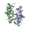
Mass: 92.094 Da / Num. of mol.: 4 / Source method: obtained synthetically / Formula: C3H8O3
#4: Water ChemComp-HOH / water


Mass: 18.015 Da / Num. of mol.: 468 / Source method: isolated from a natural source / Formula: H2O

-
Experimental details

-
Experiment

ExperimentMethod: X-RAY DIFFRACTION / Number of used crystals: 1

-
Sample preparation

CrystalDensity Matthews: 2.4 Å3/Da / Density % sol: 48.69 %
Crystal growTemperature: 298 K / Method: vapor diffusion / pH: 8
Details: PEG 8000, HEPES, pH 8.0, vapor diffusion, temperature 298K

-
Data collection

DiffractionMean temperature: 100 K
Diffraction sourceSource: SYNCHROTRON / Site: ALS / Beamline: 8.3.1 / Wavelength: 1.115872 Å
DetectorType: ADSC QUANTUM 315r / Detector: CCD / Date: Dec 13, 2008
RadiationMonochromator: Double flat crystal Si(111) / Protocol: SINGLE WAVELENGTH / Monochromatic (M) / Laue (L): M / Scattering type: x-ray
Radiation wavelengthWavelength: 1.115872 Å / Relative weight: 1
ReflectionResolution: 1.33→50 Å / Num. obs: 97906 / % possible obs: 98.3 % / Redundancy: 3.8 % / Rmerge(I) obs: 0.05 / Net I/σ(I): 16.9
Reflection shell
Resolution (Å)Redundancy (%)Rmerge(I) obs% possible all
1.33-1.383.70.48995.7
1.38-1.433.90.37997.3
1.43-1.53.90.27397.9
1.5-1.583.90.19998.4
1.58-1.683.90.1598.6
1.68-1.813.90.10999.1
1.81-1.993.80.07699.3
1.99-2.273.70.05799.3
2.27-2.873.70.04699.3
2.87-503.80.03698.1

-
Phasing

PhasingMethod: molecular replacement
Phasing MRRfactor: 55.26 / Model details: Phaser MODE: MR_AUTO
Highest resolutionLowest resolution
Rotation2.5 Å42.49 Å
Translation2.5 Å42.49 Å

-
Processing

Software
NameVersionClassificationNB
DENZOdata reduction
SCALEPACKdata scaling
PHASER2.1.1phasing
REFMAC5.5.0066refinement
PDB_EXTRACT3.005data extraction
HKL-2000data collection
HKL-2000data reduction
HKL-2000data scaling
RefinementMethod to determine structure: MOLECULAR REPLACEMENT / Resolution: 1.33→40.95 Å / Cor.coef. Fo:Fc: 0.956 / Cor.coef. Fo:Fc free: 0.954 / Occupancy max: 1 / Occupancy min: 0.3 / SU B: 1.444 / SU ML: 0.028 / Cross valid method: THROUGHOUT / ESU R: 0.061 / ESU R Free: 0.055 / Stereochemistry target values: MAXIMUM LIKELIHOOD / Details: HYDROGENS HAVE BEEN ADDED IN THE RIDING POSITIONS
RfactorNum. reflection% reflectionSelection details
Rfree0.20211 4871 5 %RANDOM
Rwork0.18199 ---
obs0.18298 92958 98.25 %-
Solvent computationIon probe radii: 0.8 Å / Shrinkage radii: 0.8 Å / VDW probe radii: 1.2 Å / Solvent model: MASK
Displacement parametersBiso mean: 15.082 Å2
Baniso -1Baniso -2Baniso -3
1-0.16 Å20 Å20 Å2
2--0.37 Å20 Å2
3----0.53 Å2
Refinement stepCycle: LAST / Resolution: 1.33→40.95 Å
ProteinNucleic acidLigandSolventTotal
Num. atoms3105 0 24 468 3597
Refine LS restraints
Refine-IDTypeDev idealDev ideal targetNumber
X-RAY DIFFRACTIONr_bond_refined_d0.0080.0223305
X-RAY DIFFRACTIONr_bond_other_d
X-RAY DIFFRACTIONr_angle_refined_deg1.3062.014510
X-RAY DIFFRACTIONr_angle_other_deg
X-RAY DIFFRACTIONr_dihedral_angle_1_deg5.3825423
X-RAY DIFFRACTIONr_dihedral_angle_2_deg31.45522.794136
X-RAY DIFFRACTIONr_dihedral_angle_3_deg10.84815.055545
X-RAY DIFFRACTIONr_dihedral_angle_4_deg17.9771530
X-RAY DIFFRACTIONr_chiral_restr0.0780.2497
X-RAY DIFFRACTIONr_gen_planes_refined0.0080.0222544
X-RAY DIFFRACTIONr_gen_planes_other
X-RAY DIFFRACTIONr_nbd_refined
X-RAY DIFFRACTIONr_nbd_other
X-RAY DIFFRACTIONr_nbtor_refined
X-RAY DIFFRACTIONr_nbtor_other
X-RAY DIFFRACTIONr_xyhbond_nbd_refined
X-RAY DIFFRACTIONr_xyhbond_nbd_other
X-RAY DIFFRACTIONr_metal_ion_refined
X-RAY DIFFRACTIONr_metal_ion_other
X-RAY DIFFRACTIONr_symmetry_vdw_refined
X-RAY DIFFRACTIONr_symmetry_vdw_other
X-RAY DIFFRACTIONr_symmetry_hbond_refined
X-RAY DIFFRACTIONr_symmetry_hbond_other
X-RAY DIFFRACTIONr_symmetry_metal_ion_refined
X-RAY DIFFRACTIONr_symmetry_metal_ion_other
X-RAY DIFFRACTIONr_mcbond_it1.0621.52074
X-RAY DIFFRACTIONr_mcbond_other
X-RAY DIFFRACTIONr_mcangle_it1.72723382
X-RAY DIFFRACTIONr_scbond_it2.41331231
X-RAY DIFFRACTIONr_scangle_it3.7284.51123
X-RAY DIFFRACTIONr_rigid_bond_restr1.27733305
X-RAY DIFFRACTIONr_sphericity_free3.8253468
X-RAY DIFFRACTIONr_sphericity_bonded2.83133220
LS refinement shellResolution: 1.329→1.363 Å / Total num. of bins used: 20
RfactorNum. reflection% reflection
Rfree0.37 336 -
Rwork0.336 6513 -
obs--94.31 %
Refinement TLS params.

Method: refined / Refine-ID: X-RAY DIFFRACTION

IDL112)L122)L132)L222)L232)L332)S11 (Å °)S12 (Å °)S13 (Å °)S21 (Å °)S22 (Å °)S23 (Å °)S31 (Å °)S32 (Å °)S33 (Å °)T112)T122)T132)T222)T232)T332)Origin x (Å)Origin y (Å)Origin z (Å)
10.4983-0.20720.31011.6615-0.51180.87150.0113-0.04130.0145-0.0001-0.0262-0.015-0.00420.02970.01490.0005-0.00250.00060.0206-0.00620.003211.3517-6.3348-18.9133
20.48910.4676-0.02560.9018-0.01710.34290.00950.0259-0.03950.01310.0091-0.02420.00660.0232-0.01870.00440.00770.00250.01790.00050.008618.0702-23.8535-42.3369
31.5115-0.3908-0.2462.5233-0.33593.61-0.01690.0132-0.1668-0.1086-0.01890.01110.1471-0.12690.03580.04710.0010.00290.056-0.00430.05311.0228-11.4171-29.6613
44.95321.3756-1.88159.9675-3.99394.27030.0037-0.095-0.09230.29480.12850.31340.0329-0.0172-0.13220.0272-0.00310.01740.0109-0.0010.024712.9551-19.979-33.3908
Refinement TLS group
IDRefine-IDRefine TLS-IDAuth asym-IDAuth seq-ID
1X-RAY DIFFRACTION1A1892 - 2085
2X-RAY DIFFRACTION2B1892 - 2085
3X-RAY DIFFRACTION3C139 - 142
4X-RAY DIFFRACTION4D139 - 142

+
About Yorodumi

-
News

-
Feb 9, 2022. New format data for meta-information of EMDB entries

New format data for meta-information of EMDB entries

  • Version 3 of the EMDB header file is now the official format.
  • The previous official version 1.9 will be removed from the archive.

Related info.:EMDB header

External links:wwPDB to switch to version 3 of the EMDB data model

-
Aug 12, 2020. Covid-19 info

Covid-19 info

URL: https://pdbjlvh1.pdbj.org/emnavi/covid19.php

New page: Covid-19 featured information page in EM Navigator.

Related info.:Covid-19 info / Mar 5, 2020. Novel coronavirus structure data

+
Mar 5, 2020. Novel coronavirus structure data

Novel coronavirus structure data

Related info.:Yorodumi Speices / Aug 12, 2020. Covid-19 info

External links:COVID-19 featured content - PDBj / Molecule of the Month (242):Coronavirus Proteases

+
Jan 31, 2019. EMDB accession codes are about to change! (news from PDBe EMDB page)

EMDB accession codes are about to change! (news from PDBe EMDB page)

  • The allocation of 4 digits for EMDB accession codes will soon come to an end. Whilst these codes will remain in use, new EMDB accession codes will include an additional digit and will expand incrementally as the available range of codes is exhausted. The current 4-digit format prefixed with “EMD-” (i.e. EMD-XXXX) will advance to a 5-digit format (i.e. EMD-XXXXX), and so on. It is currently estimated that the 4-digit codes will be depleted around Spring 2019, at which point the 5-digit format will come into force.
  • The EM Navigator/Yorodumi systems omit the EMD- prefix.

Related info.:Q: What is EMD? / ID/Accession-code notation in Yorodumi/EM Navigator

External links:EMDB Accession Codes are Changing Soon! / Contact to PDBj

+
Jul 12, 2017. Major update of PDB

Major update of PDB

  • wwPDB released updated PDB data conforming to the new PDBx/mmCIF dictionary.
  • This is a major update changing the version number from 4 to 5, and with Remediation, in which all the entries are updated.
  • In this update, many items about electron microscopy experimental information are reorganized (e.g. em_software).
  • Now, EM Navigator and Yorodumi are based on the updated data.

External links:wwPDB Remediation / Enriched Model Files Conforming to OneDep Data Standards Now Available in the PDB FTP Archive

-
Yorodumi

Thousand views of thousand structures

  • Yorodumi is a browser for structure data from EMDB, PDB, SASBDB, etc.
  • This page is also the successor to EM Navigator detail page, and also detail information page/front-end page for Omokage search.
  • The word "yorodu" (or yorozu) is an old Japanese word meaning "ten thousand". "mi" (miru) is to see.

Related info.:EMDB / PDB / SASBDB / Comparison of 3 databanks / Yorodumi Search / Aug 31, 2016. New EM Navigator & Yorodumi / Yorodumi Papers / Jmol/JSmol / Function and homology information / Changes in new EM Navigator and Yorodumi

Read more