 Movie
Movie Controller

[English] 日本語
 Yorodumi
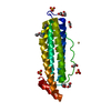
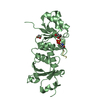
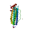
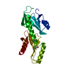
- PDB-2lm5: Solution structure of Ca2+-CIB1 in complex with the cytoplasmic d... -
+ Open data

- Basic information
| Entry | Database: PDB / ID: 2lm5 | ||||||
|---|---|---|---|---|---|---|---|
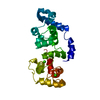
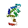
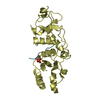
| Title | Solution structure of Ca2+-CIB1 in complex with the cytoplasmic domain of the integrin aIIb subunit | ||||||
 Components Components | Calcium and integrin-binding protein 1 | ||||||
 Keywords Keywords | METAL BINDING PROTEIN | ||||||
| Function / homology |  Function and homology information Function and homology informationpositive regulation of male germ cell proliferation / calcium-dependent protein kinase inhibitor activity / endomitotic cell cycle / positive regulation of catalytic activity / filopodium tip / thrombopoietin-mediated signaling pathway / negative regulation of microtubule depolymerization / positive regulation of calcineurin-NFAT signaling cascade / positive regulation of cell adhesion mediated by integrin / platelet formation ...positive regulation of male germ cell proliferation / calcium-dependent protein kinase inhibitor activity / endomitotic cell cycle / positive regulation of catalytic activity / filopodium tip / thrombopoietin-mediated signaling pathway / negative regulation of microtubule depolymerization / positive regulation of calcineurin-NFAT signaling cascade / positive regulation of cell adhesion mediated by integrin / platelet formation / positive regulation of cell migration involved in sprouting angiogenesis / positive regulation of cell-matrix adhesion / negative regulation of protein phosphorylation / positive regulation of protein serine/threonine kinase activity / spermatid development / regulation of cell division / positive regulation of protein targeting to membrane / protein serine/threonine kinase inhibitor activity / negative regulation of megakaryocyte differentiation / extrinsic apoptotic signaling pathway / cytoplasmic microtubule organization / protein-membrane adaptor activity / positive regulation of substrate adhesion-dependent cell spreading / response to ischemia / negative regulation of phosphatidylinositol 3-kinase/protein kinase B signal transduction / : / cell periphery / positive regulation of protein localization to plasma membrane / cellular response to nerve growth factor stimulus / sarcolemma / positive regulation of protein phosphorylation / cellular response to growth factor stimulus / cellular response to tumor necrosis factor / small GTPase binding / ruffle membrane / double-strand break repair / regulation of cell population proliferation / lamellipodium / negative regulation of neuron projection development / growth cone / positive regulation of cell growth / angiogenesis / vesicle / transmembrane transporter binding / perikaryon / positive regulation of ERK1 and ERK2 cascade / cell adhesion / apical plasma membrane / neuron projection / nuclear body / positive regulation of cell migration / negative regulation of cell population proliferation / axon / cell division / neuronal cell body / apoptotic process / calcium ion binding / positive regulation of cell population proliferation / DNA damage response / centrosome / negative regulation of apoptotic process / perinuclear region of cytoplasm / magnesium ion binding / Golgi apparatus / endoplasmic reticulum / extracellular exosome / nucleoplasm / membrane / nucleus / plasma membrane / cytoplasm Similarity search - Function | ||||||
| Biological species |  Homo sapiens (human) Homo sapiens (human) | ||||||
| Method | SOLUTION NMR / simulated annealing | ||||||
| Model details | lowest energy, model 2 | ||||||
 Authors Authors | Huang, H. / Vogel, H.J. | ||||||
 Citation Citation | Journal: J.Am.Chem.Soc. / Year: 2012 Title: Structural basis for the activation of platelet integrin alphaIIb-beta3 by calcium- and integrin-binding protein 1. Authors: Huang, H. / Vogel, H.J. | ||||||
| History |
|
- Structure visualization
| Structure viewer | Molecule: MolmilJmol/JSmol |
|---|
- Downloads & links
-Download
| PDBx/mmCIF format | 2lm5.cif.gz | 580.3 KB | Display | PDBx/mmCIF format |
|---|---|---|---|---|
| PDB format | pdb2lm5.ent.gz | 479.5 KB | Display | PDB format |
| PDBx/mmJSON format | 2lm5.json.gz | Tree view | PDBx/mmJSON format | |
| Others |  Other downloads Other downloads |
-Validation report
| Arichive directory | https://data.pdbj.org/pub/pdb/validation_reports/lm/2lm5ftp://data.pdbj.org/pub/pdb/validation_reports/lm/2lm5 | HTTPS FTP |
|---|
-Related structure data
| Related structure data | |
|---|---|
| Similar structure data | |
| Other databases |


-Links
 PDBj
PDBj- Assembly
| Deposited unit | 
| |||||||||
|---|---|---|---|---|---|---|---|---|---|---|
| 1 |
| |||||||||
| NMR ensembles |
|
-Components
| #1: Protein | Mass: 24507.264 Da / Num. of mol.: 1 Source method: isolated from a genetically manipulated source Source: (gene. exp.) Homo sapiens (human) / Gene: CIB1, CIB, KIP, PRKDCIP / Production host:  |
|---|---|
| #2: Chemical |   Mass: 40.078 Da / Num. of mol.: 2 / Source method: obtained synthetically / Formula: Ca Mass: 40.078 Da / Num. of mol.: 2 / Source method: obtained synthetically / Formula: Ca |
-Experimental details
-Experiment
| Experiment | Method: SOLUTION NMR | ||||||||||||||||||||||||||||||||||||||||
|---|---|---|---|---|---|---|---|---|---|---|---|---|---|---|---|---|---|---|---|---|---|---|---|---|---|---|---|---|---|---|---|---|---|---|---|---|---|---|---|---|---|
| NMR experiment |
|
 HSQC
HSQC- Sample preparation
| Details |
| |||||||||||||||||||||||||||||||||||||||||||||||||||||||||||||||||||||||||||||||||||||||||||||||||||||||||||||||||||||||||||||||||||||||||||||||||||||
|---|---|---|---|---|---|---|---|---|---|---|---|---|---|---|---|---|---|---|---|---|---|---|---|---|---|---|---|---|---|---|---|---|---|---|---|---|---|---|---|---|---|---|---|---|---|---|---|---|---|---|---|---|---|---|---|---|---|---|---|---|---|---|---|---|---|---|---|---|---|---|---|---|---|---|---|---|---|---|---|---|---|---|---|---|---|---|---|---|---|---|---|---|---|---|---|---|---|---|---|---|---|---|---|---|---|---|---|---|---|---|---|---|---|---|---|---|---|---|---|---|---|---|---|---|---|---|---|---|---|---|---|---|---|---|---|---|---|---|---|---|---|---|---|---|---|---|---|---|---|---|
| Sample |
|