Monochromator: Side-scattering cuberoot I-beam bent single crystal; asymetric cut 12.2 degs. Protocol: SINGLE WAVELENGTH / Monochromatic (M) / Laue (L): M / Scattering type: x-ray
Radiation wavelength
Wavelength: 0.97908 Å / Relative weight: 1
Reflection
Redundancy: 6.9 % / Av σ(I) over netI: 25.1 / Number: 108109 / Rmerge(I) obs: 0.07 / Χ2: 1 / D res high: 2.7 Å / D res low: 35 Å / Num. obs: 15594 / % possible obs: 92.2
Diffraction reflection shell
Highest resolution (Å)
Lowest resolution (Å)
% possible obs (%)
ID
Rmerge(I) obs
Chi squared
Redundancy
5.81
35
93.5
1
0.038
0.867
8.1
4.61
5.81
95
1
0.057
0.98
8.1
4.03
4.61
95.5
1
0.056
0.991
8
3.66
4.03
96
1
0.073
1.052
8.1
3.4
3.66
96
1
0.099
1.005
8
3.2
3.4
96.2
1
0.14
0.993
7.7
3.04
3.2
95
1
0.202
1.047
7
2.91
3.04
92.8
1
0.267
1.103
5.6
2.8
2.91
87
1
0.29
1.086
4.3
2.7
2.8
74.5
1
0.296
1.028
3.5
Reflection
Resolution: 2.7→35 Å / Num. obs: 15594 / % possible obs: 92.2 % / Redundancy: 6.9 % / Rmerge(I) obs: 0.07 / Χ2: 1.005 / Net I/σ(I): 14.5
Reflection shell
Resolution (Å)
Redundancy (%)
Rmerge(I) obs
Num. unique all
Χ2
Diffraction-ID
% possible all
2.7-2.8
3.5
0.296
1259
1.028
1
74.5
2.8-2.91
4.3
0.29
1483
1.086
1
87
2.91-3.04
5.6
0.267
1540
1.103
1
92.8
3.04-3.2
7
0.202
1622
1.047
1
95
3.2-3.4
7.7
0.14
1630
0.993
1
96.2
3.4-3.66
8
0.099
1636
1.005
1
96
3.66-4.03
8.1
0.073
1601
1.052
1
96
4.03-4.61
8
0.056
1609
0.991
1
95.5
4.61-5.81
8.1
0.057
1626
0.98
1
95
5.81-35
8.1
0.038
1588
0.867
1
93.5
-
Processing
Software
Name
Version
Classification
NB
DENZO
datareduction
SCALEPACK
datascaling
PHENIX
1.6.4_486
refinement
PDB_EXTRACT
3.1
dataextraction
HKL-2000
datacollection
PHENIX
phasing
Refinement
Method to determine structure: SAD / Resolution: 2.702→31.784 Å / Occupancy max: 1 / Occupancy min: 1 / SU ML: 0.37 / σ(F): 0 / Phase error: 25.32 / Stereochemistry target values: MLHL
Rfactor
Num. reflection
% reflection
Selection details
Rfree
0.2544
847
10.09 %
RANDOM
Rwork
0.2067
-
-
-
obs
0.2116
8392
91.4 %
-
all
-
9239
-
-
Solvent computation
Shrinkage radii: 0.89 Å / VDW probe radii: 1 Å / Solvent model: FLAT BULK SOLVENT MODEL / Bsol: 44.395 Å2 / ksol: 0.39 e/Å3
 Movie
Movie Controller

 Yorodumi
 Open data
 Basic information
 Components
 Keywords
 Function and homology information
 X-RAY DIFFRACTION /
  Authors
 Citation
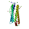
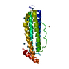
 Structure visualization
 Downloads & links
 Other downloads


 PDBj
PDBj
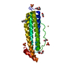
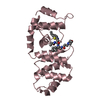
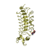
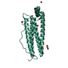
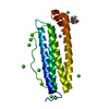
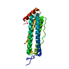
 Assembly


 Mass: 96.063 Da / Num. of mol.: 1 / Source method: obtained synthetically / Formula: SO4
 Mass: 18.015 Da / Num. of mol.: 31 / Source method: isolated from a natural source / Formula: H2O
 Sample preparation
 / Beamline: BL9-1 / Wavelength: 0.97908 Å
 Processing