 Movie
Movie Controller


+ Open data

- Basic information
| Entry | Database: PDB / ID: 6gkb | ||||||
|---|---|---|---|---|---|---|---|
| Title | Iron soak structure of Y40F SynFtn | ||||||
 Components Components | Ferritin | ||||||
 Keywords Keywords | METAL BINDING PROTEIN / Ferritin | ||||||
| Function / homology |  Function and homology information Function and homology informationbacterial non-heme ferritin / ferric iron binding / iron ion transport / ferrous iron binding / intracellular iron ion homeostasis / cytoplasm Similarity search - Function | ||||||
| Biological species |  Synechococcus sp. (bacteria) Synechococcus sp. (bacteria) | ||||||
| Method |  X-RAY DIFFRACTION / SYNCHROTRON / MOLECULAR REPLACEMENT / Resolution: 1.9 Å X-RAY DIFFRACTION / SYNCHROTRON / MOLECULAR REPLACEMENT / Resolution: 1.9 Å | ||||||
 Authors Authors | Hemmings, A.M. / Bradley, J.M. | ||||||
| Funding support |  United Kingdom, 1items United Kingdom, 1items
| ||||||
 Citation Citation | Journal: Proc. Natl. Acad. Sci. U.S.A. / Year: 2019 Title: Reaction of O2with a diiron protein generates a mixed-valent Fe2+/Fe3+center and peroxide. Authors: Bradley, J.M. / Svistunenko, D.A. / Pullin, J. / Hill, N. / Stuart, R.K. / Palenik, B. / Wilson, M.T. / Hemmings, A.M. / Moore, G.R. / Le Brun, N.E. | ||||||
| History |
|
- Structure visualization
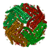
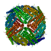
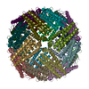
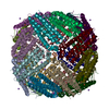
| Structure viewer | Molecule: MolmilJmol/JSmol |
|---|
- Downloads & links
-Download
| PDBx/mmCIF format | 6gkb.cif.gz | 91.4 KB | Display | PDBx/mmCIF format |
|---|---|---|---|---|
| PDB format | pdb6gkb.ent.gz | 70 KB | Display | PDB format |
| PDBx/mmJSON format | 6gkb.json.gz | Tree view | PDBx/mmJSON format | |
| Others |  Other downloads Other downloads |
-Validation report
| Arichive directory | https://data.pdbj.org/pub/pdb/validation_reports/gk/6gkbftp://data.pdbj.org/pub/pdb/validation_reports/gk/6gkb | HTTPS FTP |
|---|
-Related structure data
| Related structure data | 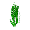 5ouwC 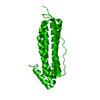 5ouzC 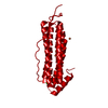 6gkaC 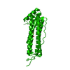 6gkcC  1eumS C: citing same article ( S: Starting model for refinement |
|---|---|
| Similar structure data |


-Links
 PDBj
PDBj


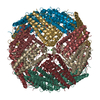
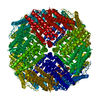
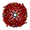
- Assembly
| Deposited unit | 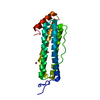
| |||||||||||||||
|---|---|---|---|---|---|---|---|---|---|---|---|---|---|---|---|---|
| 1 | x 24
| |||||||||||||||
| Unit cell |
| |||||||||||||||
| Components on special symmetry positions |
|
-Components
| #1: Protein | Mass: 19797.893 Da / Num. of mol.: 1 Source method: isolated from a genetically manipulated source Source: (gene. exp.) Synechococcus sp. (strain CC9311) (bacteria)Strain: CC9311 / Gene: sync_1539 / Production host: | ||
|---|---|---|---|
| #2: Chemical |   Mass: 55.845 Da / Num. of mol.: 2 / Source method: obtained synthetically / Formula: Fe / Feature type: SUBJECT OF INVESTIGATION Mass: 55.845 Da / Num. of mol.: 2 / Source method: obtained synthetically / Formula: Fe / Feature type: SUBJECT OF INVESTIGATION#3: Water | ChemComp-HOH / |  Mass: 18.015 Da / Num. of mol.: 153 / Source method: isolated from a natural source / Formula: H2O Mass: 18.015 Da / Num. of mol.: 153 / Source method: isolated from a natural source / Formula: H2O |
-Experimental details
-Experiment
| Experiment | Method: X-RAY DIFFRACTION / Number of used crystals: 1 |
|---|
- Sample preparation
| Crystal | Density Matthews: 2.9 Å3/Da / Density % sol: 57.61 % |
|---|---|
| Crystal grow | Temperature: 289 K / Method: vapor diffusion, sitting drop / pH: 4.6 / Details: 0.1 M sodium acetate 2.0 M sodium chloride |
-Data collection
| Diffraction | Mean temperature: 100 K |
|---|---|
| Diffraction source | Source: SYNCHROTRON / Site: Diamond / Beamline: I04 / Wavelength: 0.97949 Å |
| Detector | Type: DECTRIS PILATUS 6M-F / Detector: PIXEL / Date: Nov 20, 2014 |
| Radiation | Protocol: SINGLE WAVELENGTH / Monochromatic (M) / Laue (L): M / Scattering type: x-ray |
| Radiation wavelength | Wavelength: 0.97949 Å / Relative weight: 1 |
| Reflection | Resolution: 1.9→62.47 Å / Num. obs: 18124 / % possible obs: 95.9 % / Redundancy: 4.1 % / Rmerge(I) obs: 0.047 / Rpim(I) all: 0.036 / Rrim(I) all: 0.059 / Net I/σ(I): 14.4 |
| Reflection shell | Resolution: 1.9→1.94 Å / Redundancy: 4 % / Rmerge(I) obs: 0.855 / Mean I/σ(I) obs: 1.4 / Num. unique obs: 1149 / Rpim(I) all: 0.648 / Rrim(I) all: 1.078 / % possible all: 98.1 |
- Processing
| Software |
| |||||||||||||||||||||||||||||||||||||||||||||||||
|---|---|---|---|---|---|---|---|---|---|---|---|---|---|---|---|---|---|---|---|---|---|---|---|---|---|---|---|---|---|---|---|---|---|---|---|---|---|---|---|---|---|---|---|---|---|---|---|---|---|---|
| Refinement | Method to determine structure: MOLECULAR REPLACEMENT Starting model: 1EUM Resolution: 1.9→62.465 Å / SU ML: 0.21 / Cross valid method: THROUGHOUT / σ(F): 1.34 / Phase error: 20.27
| |||||||||||||||||||||||||||||||||||||||||||||||||
| Solvent computation | Shrinkage radii: 0.9 Å / VDW probe radii: 1.11 Å | |||||||||||||||||||||||||||||||||||||||||||||||||
| Displacement parameters | Biso max: 81.05 Å2 / Biso mean: 39.4616 Å2 / Biso min: 24.81 Å2 | |||||||||||||||||||||||||||||||||||||||||||||||||
| Refinement step | Cycle: final / Resolution: 1.9→62.465 Å
| |||||||||||||||||||||||||||||||||||||||||||||||||
| LS refinement shell | Refine-ID: X-RAY DIFFRACTION / Rfactor Rfree error: 0 / Total num. of bins used: 6
| |||||||||||||||||||||||||||||||||||||||||||||||||
| Refinement TLS params. | Method: refined / Origin x: 50.6379 Å / Origin y: 187.0773 Å / Origin z: 205.1403 Å
| |||||||||||||||||||||||||||||||||||||||||||||||||
| Refinement TLS group |
|
