 Movie
Movie Controller


+ Open data

- Basic information
| Entry | Database: PDB / ID: 7kod | |||||||||||||||||||||
|---|---|---|---|---|---|---|---|---|---|---|---|---|---|---|---|---|---|---|---|---|---|---|
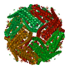
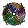
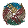
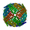
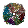
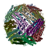
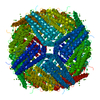
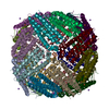
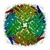
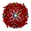
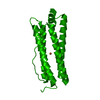
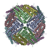
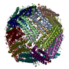
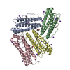
| Title | Cryo-EM structure of heavy chain mouse apoferritin | |||||||||||||||||||||
 Components Components | Ferritin heavy chain | |||||||||||||||||||||
 Keywords Keywords | OXIDOREDUCTASE / protein | |||||||||||||||||||||
| Function / homology |  Function and homology information Function and homology informationIron uptake and transport / Golgi Associated Vesicle Biogenesis / ferroxidase / negative regulation of ferroptosis / autolysosome / ferroxidase activity / negative regulation of fibroblast proliferation / endocytic vesicle lumen / Neutrophil degranulation / ferric iron binding ...Iron uptake and transport / Golgi Associated Vesicle Biogenesis / ferroxidase / negative regulation of ferroptosis / autolysosome / ferroxidase activity / negative regulation of fibroblast proliferation / endocytic vesicle lumen / Neutrophil degranulation / ferric iron binding / autophagosome / iron ion transport / ferrous iron binding / intracellular iron ion homeostasis / immune response / iron ion binding / negative regulation of cell population proliferation / mitochondrion / extracellular region / membrane / identical protein binding / cytoplasm / cytosol Similarity search - Function | |||||||||||||||||||||
| Biological species |  | |||||||||||||||||||||
| Method | ELECTRON MICROSCOPY / single particle reconstruction / cryo EM / Resolution: 1.655 Å | |||||||||||||||||||||
 Authors Authors | Sun, M. / Azumaya, C. / Tse, E. / Frost, A. / Southworth, D. / Verba, K.A. / Cheng, Y. / Agard, D.A. | |||||||||||||||||||||
| Funding support |  United States, 6items United States, 6items
| |||||||||||||||||||||
 Citation Citation | Journal: J Struct Biol / Year: 2021 Title: Practical considerations for using K3 cameras in CDS mode for high-resolution and high-throughput single particle cryo-EM. Authors: Ming Sun / Caleigh M Azumaya / Eric Tse / David P Bulkley / Matthew B Harrington / Glenn Gilbert / Adam Frost / Daniel Southworth / Kliment A Verba / Yifan Cheng / David A Agard / Abstract: Detector technology plays a pivotal role in high-resolution and high-throughput cryo-EM structure determination. Compared with the first-generation, single-electron counting direct detection camera ...Detector technology plays a pivotal role in high-resolution and high-throughput cryo-EM structure determination. Compared with the first-generation, single-electron counting direct detection camera (Gatan K2), the latest K3 camera is faster, larger, and now offers a correlated-double sampling mode (CDS). Importantly this results in a higher DQE and improved throughput compared to its predecessor. In this study, we focused on optimizing camera data collection parameters for daily use within a cryo-EM facility and explored the balance between throughput and resolution. In total, eight data sets of murine heavy-chain apoferritin were collected at different dose rates and magnifications, using 9-hole image shift data collection strategies. The performance of the camera was characterized by the quality of the resultant 3D reconstructions. Our results demonstrated that the Gatan K3 operating in CDS mode outperformed standard (nonCDS) mode in terms of reconstruction resolution in all tested conditions with 8 electrons per pixel per second being the optimal dose rate. At low magnification (64kx) we were able to achieve reconstruction resolutions of 149% of the physical Nyquist limit (1.8 Å with a 1.346 Å physical pixel size). Low magnification allows more particles to be collected per image, aiding analysis of heterogeneous samples requiring large data sets. At moderate magnification (105kx, 0.834 Å physical pixel size) we achieved a resolution of 1.65 Å within 8-h of data collection, a condition optimal for achieving high-resolution on well behaved samples. Our results also show that for an optimal sample like apoferritin, one can achieve better than 2.5 Å resolution with 5 min of data collection. Together, our studies validate the most efficient ways of imaging protein complexes using the K3 direct detector and will greatly benefit the cryo-EM community. | |||||||||||||||||||||
| History |
|
- Structure visualization
| Movie |
Movie viewer |
|---|---|
| Structure viewer | Molecule: MolmilJmol/JSmol |
- Downloads & links
-Download
| PDBx/mmCIF format | 7kod.cif.gz | 1.5 MB | Display | PDBx/mmCIF format |
|---|---|---|---|---|
| PDB format | pdb7kod.ent.gz | 1.3 MB | Display | PDB format |
| PDBx/mmJSON format | 7kod.json.gz | Tree view | PDBx/mmJSON format | |
| Others |  Other downloads Other downloads |
-Validation report
| Arichive directory | https://data.pdbj.org/pub/pdb/validation_reports/ko/7kodftp://data.pdbj.org/pub/pdb/validation_reports/ko/7kod | HTTPS FTP |
|---|
-Related structure data
| Related structure data |  22972MC M: map data used to model this data C: citing same article ( |
|---|---|
| Similar structure data | |
| EM raw data | EMPIAR-10588 (Title: Cryo-EM reconstruction of heavy chain mouse apoferritin, 8 datasets testing dose rate and correlative double sampling mode for the Gatan K3 direct electron detector. [1143 multi-frame ...Title: Cryo-EM reconstruction of heavy chain mouse apoferritin, 8 datasets testing dose rate and correlative double sampling mode for the Gatan K3 direct electron detector. [1143 multi-frame micrographs composed of 120 frames in LZW compressed TIFF format Data size: 8.5 TB Data #1: apoferritin multi-frame raw micrographs non gain normalized frames in LZW compressed TIFF format collected at 8 e/p/s in CDS mode from Gatan K3 [micrographs - multiframe] Data #2: apoferritin multi-frame raw micrographs non gain normalized frames in LZW compressed TIFF format collected at 8 e/p/s in standard mode from Gatan K3 [micrographs - multiframe] Data #3: apoferritin multi-frame raw micrographs non gain normalized frames in LZW compressed TIFF format collected at 4 e/p/s in CDS mode from Gatan K3 [micrographs - multiframe] Data #4: apoferritin multi-frame raw micrographs non gain normalized frames in LZW compressed TIFF format collected at 4 e/p/s in CDS mode from Gatan K3 [micrographs - multiframe] Data #5: apoferritin multi-frame raw micrographs non gain normalized frames in LZW compressed TIFF format collected at 8 e/p/s in CDS mode from Gatan K3 at lower magnification [micrographs - multiframe] Data #6: apoferritin multi-frame raw micrographs non gain normalized frames in LZW compressed TIFF format collected at 8 e/p/s in standard mode from Gatan K3 at lower magnification [micrographs - multiframe] Data #7: apoferritin multi-frame raw micrographs non gain normalized frames in LZW compressed TIFF format collected at 16 e/p/s in CDS mode from Gatan K3 at lower magnification [micrographs - multiframe] Data #8: apoferritin multi-frame raw micrographs non gain normalized frames in LZW compressed TIFF format collected at 16 e/p/s in standard mode from Gatan K3 at lower magnification [micrographs - multiframe]) |


-Links
 PDBj
PDBj


- Assembly
| Deposited unit | 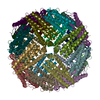
|
|---|---|
| 1 |
|
-Components
| #1: Protein | Mass: 21097.631 Da / Num. of mol.: 24 Source method: isolated from a genetically manipulated source Source: (gene. exp.)  #2: Water | ChemComp-HOH / |  Mass: 18.015 Da / Num. of mol.: 1727 / Source method: isolated from a natural source / Formula: H2O Mass: 18.015 Da / Num. of mol.: 1727 / Source method: isolated from a natural source / Formula: H2O |
|---|
-Experimental details
-Experiment
| Experiment | Method: ELECTRON MICROSCOPY |
|---|---|
| EM experiment | Aggregation state: PARTICLE / 3D reconstruction method: single particle reconstruction |
- Sample preparation
| Component | Name: Heavy chain mouse apoferritin / Type: COMPLEX / Entity ID: #1 / Source: RECOMBINANT |
|---|---|
| Molecular weight | Experimental value: NO |
| Source (natural) | Organism: |
| Source (recombinant) | Organism: |
| Buffer solution | pH: 7.5 |
| Specimen | Embedding applied: NO / Shadowing applied: NO / Staining applied: NO / Vitrification applied: YES |
| Specimen support | Grid material: COPPER / Grid mesh size: 300 divisions/in. / Grid type: Quantifoil |
| Vitrification | Cryogen name: ETHANE |
- Electron microscopy imaging
| Experimental equipment |  Model: Titan Krios / Image courtesy: FEI Company |
|---|---|
| Microscopy | Model: FEI TITAN KRIOS |
| Electron gun | Electron source:  FIELD EMISSION GUN / Accelerating voltage: 300 kV / Illumination mode: FLOOD BEAM FIELD EMISSION GUN / Accelerating voltage: 300 kV / Illumination mode: FLOOD BEAM |
| Electron lens | Mode: BRIGHT FIELD |
| Specimen holder | Specimen holder model: FEI TITAN KRIOS AUTOGRID HOLDER |
| Image recording | Average exposure time: 6 sec. / Electron dose: 69 e/Å2 / Film or detector model: GATAN K3 BIOQUANTUM (6k x 4k) / Num. of grids imaged: 1 / Details: 9-hole image-shift data collection strategy |
- Processing
| Software | Name: PHENIX / Version: 1.18.2_3874: / Classification: refinement | ||||||||||||||||||||||||||||||||||||||||||||
|---|---|---|---|---|---|---|---|---|---|---|---|---|---|---|---|---|---|---|---|---|---|---|---|---|---|---|---|---|---|---|---|---|---|---|---|---|---|---|---|---|---|---|---|---|---|
| EM software |
| ||||||||||||||||||||||||||||||||||||||||||||
| CTF correction | Type: NONE | ||||||||||||||||||||||||||||||||||||||||||||
| Symmetry | Point symmetry: O (octahedral) | ||||||||||||||||||||||||||||||||||||||||||||
| 3D reconstruction | Resolution: 1.655 Å / Resolution method: FSC 0.143 CUT-OFF / Num. of particles: 571000 / Symmetry type: POINT | ||||||||||||||||||||||||||||||||||||||||||||
| Atomic model building | Protocol: FLEXIBLE FIT | ||||||||||||||||||||||||||||||||||||||||||||
| Atomic model building | PDB-ID: 5OBA Accession code: 5OBA / Source name: PDB / Type: experimental model | ||||||||||||||||||||||||||||||||||||||||||||
| Refinement | Cross valid method: NONE Stereochemistry target values: GeoStd + Monomer Library + CDL v1.2 | ||||||||||||||||||||||||||||||||||||||||||||
| Displacement parameters | Biso mean: 21.44 Å2 | ||||||||||||||||||||||||||||||||||||||||||||
| Refine LS restraints |
|

