 Movie
Movie Controller


+ Open data

- Basic information
| Entry | Database: EMDB / ID: EMD-22972 | |||||||||||||||||||||
|---|---|---|---|---|---|---|---|---|---|---|---|---|---|---|---|---|---|---|---|---|---|---|
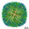
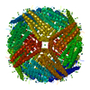
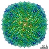
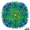
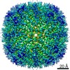
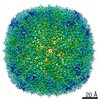
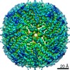
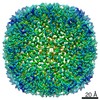
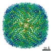
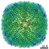
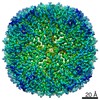
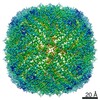
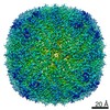
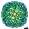
| Title | Cryo-EM structure of heavy chain mouse apoferritin | |||||||||||||||||||||
 Map data Map data | raw unsharpened map | |||||||||||||||||||||
 Sample Sample |
| |||||||||||||||||||||
 Keywords Keywords | protein / OXIDOREDUCTASE | |||||||||||||||||||||
| Function / homology |  Function and homology information Function and homology informationIron uptake and transport / Golgi Associated Vesicle Biogenesis / ferroxidase / negative regulation of ferroptosis / autolysosome / ferroxidase activity / negative regulation of fibroblast proliferation / endocytic vesicle lumen / Neutrophil degranulation / ferric iron binding ...Iron uptake and transport / Golgi Associated Vesicle Biogenesis / ferroxidase / negative regulation of ferroptosis / autolysosome / ferroxidase activity / negative regulation of fibroblast proliferation / endocytic vesicle lumen / Neutrophil degranulation / ferric iron binding / autophagosome / iron ion transport / ferrous iron binding / intracellular iron ion homeostasis / immune response / iron ion binding / negative regulation of cell population proliferation / mitochondrion / extracellular region / membrane / identical protein binding / cytoplasm / cytosol Similarity search - Function | |||||||||||||||||||||
| Biological species |  | |||||||||||||||||||||
| Method | single particle reconstruction / cryo EM / Resolution: 1.655 Å | |||||||||||||||||||||
 Authors Authors | Sun M / Azumaya C | |||||||||||||||||||||
| Funding support |  United States, 6 items United States, 6 items
| |||||||||||||||||||||
 Citation Citation | Journal: J Struct Biol / Year: 2021 Title: Practical considerations for using K3 cameras in CDS mode for high-resolution and high-throughput single particle cryo-EM. Authors: Ming Sun / Caleigh M Azumaya / Eric Tse / David P Bulkley / Matthew B Harrington / Glenn Gilbert / Adam Frost / Daniel Southworth / Kliment A Verba / Yifan Cheng / David A Agard / Abstract: Detector technology plays a pivotal role in high-resolution and high-throughput cryo-EM structure determination. Compared with the first-generation, single-electron counting direct detection camera ...Detector technology plays a pivotal role in high-resolution and high-throughput cryo-EM structure determination. Compared with the first-generation, single-electron counting direct detection camera (Gatan K2), the latest K3 camera is faster, larger, and now offers a correlated-double sampling mode (CDS). Importantly this results in a higher DQE and improved throughput compared to its predecessor. In this study, we focused on optimizing camera data collection parameters for daily use within a cryo-EM facility and explored the balance between throughput and resolution. In total, eight data sets of murine heavy-chain apoferritin were collected at different dose rates and magnifications, using 9-hole image shift data collection strategies. The performance of the camera was characterized by the quality of the resultant 3D reconstructions. Our results demonstrated that the Gatan K3 operating in CDS mode outperformed standard (nonCDS) mode in terms of reconstruction resolution in all tested conditions with 8 electrons per pixel per second being the optimal dose rate. At low magnification (64kx) we were able to achieve reconstruction resolutions of 149% of the physical Nyquist limit (1.8 Å with a 1.346 Å physical pixel size). Low magnification allows more particles to be collected per image, aiding analysis of heterogeneous samples requiring large data sets. At moderate magnification (105kx, 0.834 Å physical pixel size) we achieved a resolution of 1.65 Å within 8-h of data collection, a condition optimal for achieving high-resolution on well behaved samples. Our results also show that for an optimal sample like apoferritin, one can achieve better than 2.5 Å resolution with 5 min of data collection. Together, our studies validate the most efficient ways of imaging protein complexes using the K3 direct detector and will greatly benefit the cryo-EM community. | |||||||||||||||||||||
| History |
|
- Structure visualization
| Movie |
Movie viewer |
|---|---|
| Structure viewer | EM map: SurfViewMolmilJmol/JSmol |
| Supplemental images |

- Downloads & links
-EMDB archive
| Map data | emd_22972.map.gz | 455.6 MB | EMDB map data format | |
|---|---|---|---|---|
| Header (meta data) | emd-22972-v30.xmlemd-22972.xml | 17.4 KB 17.4 KB | Display Display | EMDB header |
| FSC (resolution estimation) | emd_22972_fsc.xml | 18.1 KB | Display | FSC data file |
| Images | 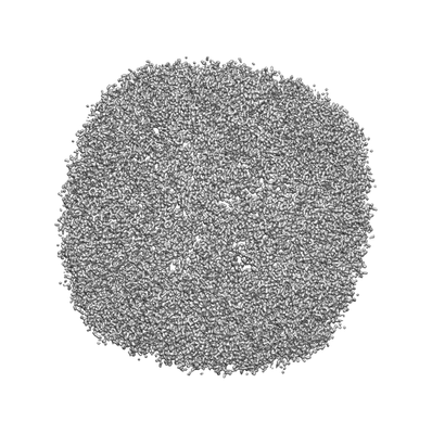 emd_22972.png emd_22972.png | 68.2 KB | ||
| Filedesc metadata | emd-22972.cif.gz | 5.6 KB | ||
| Others | emd_22972_additional_1.map.gzemd_22972_additional_2.map.gz | 76.9 MB 72.2 MB | ||
| Archive directory |  http://ftp.pdbj.org/pub/emdb/structures/EMD-22972ftp://ftp.pdbj.org/pub/emdb/structures/EMD-22972 http://ftp.pdbj.org/pub/emdb/structures/EMD-22972ftp://ftp.pdbj.org/pub/emdb/structures/EMD-22972 | HTTPS FTP |
-Related structure data
| Related structure data | 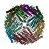 7kodMC M: atomic model generated by this map C: citing same article ( |
|---|---|
| Similar structure data | |
| EM raw data | EMPIAR-10588 (Title: Cryo-EM reconstruction of heavy chain mouse apoferritin, 8 datasets testing dose rate and correlative double sampling mode for the Gatan K3 direct electron detector. [1143 multi-frame ...Title: Cryo-EM reconstruction of heavy chain mouse apoferritin, 8 datasets testing dose rate and correlative double sampling mode for the Gatan K3 direct electron detector. [1143 multi-frame micrographs composed of 120 frames in LZW compressed TIFF format Data size: 8.5 TB Data #1: apoferritin multi-frame raw micrographs non gain normalized frames in LZW compressed TIFF format collected at 8 e/p/s in CDS mode from Gatan K3 [micrographs - multiframe] Data #2: apoferritin multi-frame raw micrographs non gain normalized frames in LZW compressed TIFF format collected at 8 e/p/s in standard mode from Gatan K3 [micrographs - multiframe] Data #3: apoferritin multi-frame raw micrographs non gain normalized frames in LZW compressed TIFF format collected at 4 e/p/s in CDS mode from Gatan K3 [micrographs - multiframe] Data #4: apoferritin multi-frame raw micrographs non gain normalized frames in LZW compressed TIFF format collected at 4 e/p/s in CDS mode from Gatan K3 [micrographs - multiframe] Data #5: apoferritin multi-frame raw micrographs non gain normalized frames in LZW compressed TIFF format collected at 8 e/p/s in CDS mode from Gatan K3 at lower magnification [micrographs - multiframe] Data #6: apoferritin multi-frame raw micrographs non gain normalized frames in LZW compressed TIFF format collected at 8 e/p/s in standard mode from Gatan K3 at lower magnification [micrographs - multiframe] Data #7: apoferritin multi-frame raw micrographs non gain normalized frames in LZW compressed TIFF format collected at 16 e/p/s in CDS mode from Gatan K3 at lower magnification [micrographs - multiframe] Data #8: apoferritin multi-frame raw micrographs non gain normalized frames in LZW compressed TIFF format collected at 16 e/p/s in standard mode from Gatan K3 at lower magnification [micrographs - multiframe]) |


-Links
| EMDB pages | EMDB (EBI/PDBe) / EMDataResource |
|---|---|
| Related items in Molecule of the Month |


-Map
| File | Download / File: emd_22972.map.gz / Format: CCP4 / Size: 512 MB / Type: IMAGE STORED AS FLOATING POINT NUMBER (4 BYTES) | ||||||||||||||||||||||||||||||||||||||||||||||||||||||||||||||||||||
|---|---|---|---|---|---|---|---|---|---|---|---|---|---|---|---|---|---|---|---|---|---|---|---|---|---|---|---|---|---|---|---|---|---|---|---|---|---|---|---|---|---|---|---|---|---|---|---|---|---|---|---|---|---|---|---|---|---|---|---|---|---|---|---|---|---|---|---|---|---|
| Annotation | raw unsharpened map | ||||||||||||||||||||||||||||||||||||||||||||||||||||||||||||||||||||
| Projections & slices | Image control
Images are generated by Spider. | ||||||||||||||||||||||||||||||||||||||||||||||||||||||||||||||||||||
| Voxel size | X=Y=Z: 0.417 Å | ||||||||||||||||||||||||||||||||||||||||||||||||||||||||||||||||||||
| Density |
| ||||||||||||||||||||||||||||||||||||||||||||||||||||||||||||||||||||
| Symmetry | Space group: 1 | ||||||||||||||||||||||||||||||||||||||||||||||||||||||||||||||||||||
| Details | EMDB XML:
CCP4 map header:
| ||||||||||||||||||||||||||||||||||||||||||||||||||||||||||||||||||||
 Z (Sec.)
 Y (Row.)
 X (Col.)


-Supplemental data
-Additional map: filtered and sharpened map (b-factor, ~ -66)
| File | emd_22972_additional_1.map | ||||||||||||
|---|---|---|---|---|---|---|---|---|---|---|---|---|---|
| Annotation | filtered and sharpened map (b-factor, ~ -66) | ||||||||||||
| Projections & Slices |
| ||||||||||||
| Density Histograms |


-Additional map: filtered and sharpened map (b-factor, ~ -25)
| File | emd_22972_additional_2.map | ||||||||||||
|---|---|---|---|---|---|---|---|---|---|---|---|---|---|
| Annotation | filtered and sharpened map (b-factor, ~ -25) | ||||||||||||
| Projections & Slices |
| ||||||||||||
| Density Histograms |


- Sample components
-Entire : Heavy chain mouse apoferritin
| Entire | Name: Heavy chain mouse apoferritin |
|---|---|
| Components |
|
-Supramolecule #1: Heavy chain mouse apoferritin
| Supramolecule | Name: Heavy chain mouse apoferritin / type: complex / ID: 1 / Parent: 0 / Macromolecule list: #1 |
|---|---|
| Source (natural) | Organism: |
-Macromolecule #1: Ferritin heavy chain
| Macromolecule | Name: Ferritin heavy chain / type: protein_or_peptide / ID: 1 / Number of copies: 24 / Enantiomer: LEVO / EC number: ferroxidase |
|---|---|
| Source (natural) | Organism: |
| Molecular weight | Theoretical: 21.097631 KDa |
| Recombinant expression | Organism:  |
| Sequence | String: MTTASPSQVR QNYHQDAEAA INRQINLELY ASYVYLSMSC YFDRDDVALK NFAKYFLHQS HEEREHAEKL MKLQNQRGGR IFLQDIKKP DRDDWESGLN AMECALHLEK SVNQSLLELH KLATDKNDPH LCDFIETYYL SEQVKSIKEL GDHVTNLRKM G APEAGMAE YLFDKHTLGH GDES UniProtKB: Ferritin heavy chain |
-Macromolecule #2: water
| Macromolecule | Name: water / type: ligand / ID: 2 / Number of copies: 1727 / Formula: HOH |
|---|---|
| Molecular weight | Theoretical: 18.015 Da |
| Chemical component information |  ChemComp-HOH: |
-Experimental details
-Structure determination
| Method | cryo EM |
|---|---|
 Processing Processing | single particle reconstruction |
| Aggregation state | particle |
-Sample preparation
| Buffer | pH: 7.5 |
|---|---|
| Grid | Model: Quantifoil / Material: COPPER / Mesh: 300 / Pretreatment - Type: GLOW DISCHARGE |
| Vitrification | Cryogen name: ETHANE |
- Electron microscopy
| Microscope | FEI TITAN KRIOS |
|---|---|
| Image recording | Film or detector model: GATAN K3 BIOQUANTUM (6k x 4k) / Number grids imaged: 1 / Average exposure time: 6.0 sec. / Average electron dose: 69.0 e/Å2 / Details: 9-hole image-shift data collection strategy |
| Electron beam | Acceleration voltage: 300 kV / Electron source:  FIELD EMISSION GUN FIELD EMISSION GUN |
| Electron optics | Illumination mode: FLOOD BEAM / Imaging mode: BRIGHT FIELD |
| Sample stage | Specimen holder model: FEI TITAN KRIOS AUTOGRID HOLDER |
| Experimental equipment |  Model: Titan Krios / Image courtesy: FEI Company |


