- EMDB-20837: Equine spleen apoferritin prepared with the Shake-it-off device -
+
Open data
ID or keywords:
Loading...
-
Basic information
Entry
Database: EMDB / ID: EMD-20837
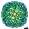
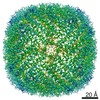
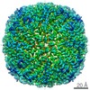
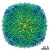
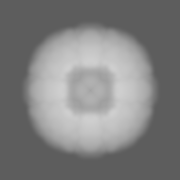
Title
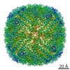
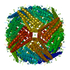
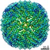
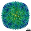
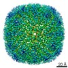
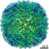
Equine spleen apoferritin prepared with the Shake-it-off device
Map data
apoferritin prepared by the shake-it-off device
Sample
Complex: apoferritin
Function / homology
Function and homology information
ferritin complex / autolysosome / ferric iron binding / autophagosome / iron ion transport / ferrous iron binding / cytoplasmic vesicle / intracellular iron ion homeostasis / iron ion binding / cytoplasm Similarity search - Function
Natural Sciences and Engineering Research Council (Canada)
Discovery Grant
Canada
Citation
Journal: Acta Crystallogr D Struct Biol / Year: 2019 Title: Shake-it-off: a simple ultrasonic cryo-EM specimen-preparation device. Authors: John L Rubinstein / Hui Guo / Zev A Ripstein / Ali Haydaroglu / Aaron Au / Christopher M Yip / Justin M Di Trani / Samir Benlekbir / Timothy Kwok / Abstract: Although microscopes and image-analysis software for electron cryomicroscopy (cryo-EM) have improved dramatically in recent years, specimen-preparation methods have lagged behind. Most strategies ...Although microscopes and image-analysis software for electron cryomicroscopy (cryo-EM) have improved dramatically in recent years, specimen-preparation methods have lagged behind. Most strategies still rely on blotting microscope grids with paper to produce a thin film of solution suitable for vitrification. This approach loses more than 99.9% of the applied sample and requires several seconds, leading to problematic air-water interface interactions for macromolecules in the resulting thin film of solution and complicating time-resolved studies. Recently developed self-wicking EM grids allow the use of small volumes of sample, with nanowires on the grid bars removing excess solution to produce a thin film within tens of milliseconds from sample application to freezing. Here, a simple cryo-EM specimen-preparation device that uses components from an ultrasonic humidifier to transfer protein solution onto a self-wicking EM grid is presented. The device is controlled by a Raspberry Pi single-board computer and all components are either widely available or can be manufactured by online services, allowing the device to be constructed in laboratories that specialize in cryo-EM rather than instrument design. The simple open-source design permits the straightforward customization of the instrument for specialized experiments.
History
Deposition
Oct 16, 2019
-
Header (metadata) release
Dec 11, 2019
-
Map release
Dec 11, 2019
-
Update
Dec 2, 2020
-
Current status
Dec 2, 2020
Processing site: RCSB / Status: Released
-
Structure visualization
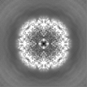
Movie
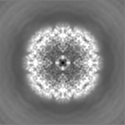
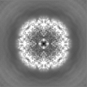
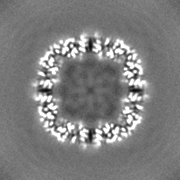
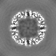
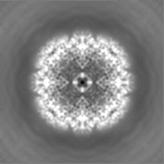
Surface view with section colored by density value
Film or detector model: FEI FALCON III (4k x 4k) / Detector mode: COUNTING / Digitization - Sampling interval: 14.0 µm / Number grids imaged: 1 / Number real images: 324 / Average exposure time: 30.0 sec. / Average electron dose: 21.0 e/Å2
Electron beam
Acceleration voltage: 300 kV / Electron source: FIELD EMISSION GUN
Electron optics
Illumination mode: FLOOD BEAM / Imaging mode: BRIGHT FIELD / Cs: 2.7 mm / Nominal magnification: 75000
In the structure databanks used in Yorodumi, some data are registered as the other names, "COVID-19 virus" and "2019-nCoV". Here are the details of the virus and the list of structure data.
Jan 31, 2019. EMDB accession codes are about to change! (news from PDBe EMDB page)
EMDB accession codes are about to change! (news from PDBe EMDB page)
The allocation of 4 digits for EMDB accession codes will soon come to an end. Whilst these codes will remain in use, new EMDB accession codes will include an additional digit and will expand incrementally as the available range of codes is exhausted. The current 4-digit format prefixed with “EMD-” (i.e. EMD-XXXX) will advance to a 5-digit format (i.e. EMD-XXXXX), and so on. It is currently estimated that the 4-digit codes will be depleted around Spring 2019, at which point the 5-digit format will come into force.
The EM Navigator/Yorodumi systems omit the EMD- prefix.
Related info.:Q: What is EMD? / ID/Accession-code notation in Yorodumi/EM Navigator
Yorodumi is a browser for structure data from EMDB, PDB, SASBDB, etc.
This page is also the successor to EM Navigator detail page, and also detail information page/front-end page for Omokage search.
The word "yorodu" (or yorozu) is an old Japanese word meaning "ten thousand". "mi" (miru) is to see.
Related info.:EMDB / PDB / SASBDB / Comparison of 3 databanks / Yorodumi Search / Aug 31, 2016. New EM Navigator & Yorodumi / Yorodumi Papers / Jmol/JSmol / Function and homology information / Changes in new EM Navigator and Yorodumi
 Movie
Movie Controller

 Yorodumi
 Open data
 Basic information
 Map data
 Sample
 Function and homology information
 Authors
 Canada, 1 items
  Citation
 Structure visualization
 Downloads & links
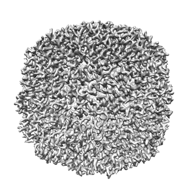 emd_20837.png
 http://ftp.pdbj.org/pub/emdb/structures/EMD-20837
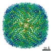


 Z (Sec.)
 Y (Row.)
 X (Col.)


 Sample components
 Processing
 Electron microscopy
 FIELD EMISSION GUN
