 Movie
Movie Controller

[English] 日本語
 Yorodumi
- PDB-3j9s: Single particle cryo-EM structure of rotavirus VP6 at 2.6 Angstro... -
+ Open data

- Basic information
| Entry | Database: PDB / ID: 3j9s | ||||||
|---|---|---|---|---|---|---|---|
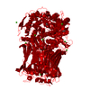
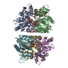
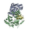
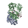
| Title | Single particle cryo-EM structure of rotavirus VP6 at 2.6 Angstrom resolution | ||||||
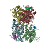
 Components Components | Intermediate capsid protein VP6 | ||||||
 Keywords Keywords | VIRAL PROTEIN / rotavirus / virus | ||||||
| Function / homology |  Function and homology information Function and homology informationviral intermediate capsid / T=13 icosahedral viral capsid / host cell surface receptor binding / fusion of virus membrane with host plasma membrane / viral envelope / structural molecule activity / metal ion binding Similarity search - Function | ||||||
| Biological species |  Bovine rotavirus strain UK/G6 Bovine rotavirus strain UK/G6 | ||||||
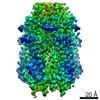
| Method | ELECTRON MICROSCOPY / single particle reconstruction / cryo EM / Resolution: 2.6 Å | ||||||
 Authors Authors | Grant, T. / Grigorieff, N. | ||||||
 Citation Citation | Journal: Elife / Year: 2015 Title: Measuring the optimal exposure for single particle cryo-EM using a 2.6 Å reconstruction of rotavirus VP6. Authors: Timothy Grant / Nikolaus Grigorieff /  Abstract: Biological specimens suffer radiation damage when imaged in an electron microscope, ultimately limiting the attainable resolution. At a given resolution, an optimal exposure can be defined that ...Biological specimens suffer radiation damage when imaged in an electron microscope, ultimately limiting the attainable resolution. At a given resolution, an optimal exposure can be defined that maximizes the signal-to-noise ratio in the image. Using a 2.6 Å resolution single particle cryo-EM reconstruction of rotavirus VP6, determined from movies recorded with a total exposure of 100 electrons/Å(2), we obtained accurate measurements of optimal exposure values over a wide range of resolutions. At low and intermediate resolutions, our measured values are considerably higher than obtained previously for crystalline specimens, indicating that both images and movies should be collected with higher exposures than are generally used. We demonstrate a method of using our optimal exposure values to filter movie frames, yielding images with improved contrast that lead to higher resolution reconstructions. This 'high-exposure' technique should benefit cryo-EM work on all types of samples, especially those of relatively low-molecular mass. | ||||||
| History |
|
- Structure visualization
| Movie |
Movie viewer |
|---|---|
| Structure viewer | Molecule: MolmilJmol/JSmol |
- Downloads & links
-Download
| PDBx/mmCIF format | 3j9s.cif.gz | 83.4 KB | Display | PDBx/mmCIF format |
|---|---|---|---|---|
| PDB format | pdb3j9s.ent.gz | 59 KB | Display | PDB format |
| PDBx/mmJSON format | 3j9s.json.gz | Tree view | PDBx/mmJSON format | |
| Others |  Other downloads Other downloads |
-Validation report
| Arichive directory | https://data.pdbj.org/pub/pdb/validation_reports/j9/3j9sftp://data.pdbj.org/pub/pdb/validation_reports/j9/3j9s | HTTPS FTP |
|---|
-Related structure data
| Related structure data | 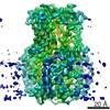 6272MC 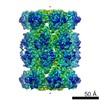 6464C M: map data used to model this data C: citing same article ( |
|---|---|
| Similar structure data |
-Links
 PDBj
PDBj


- Assembly
| Deposited unit | 
|
|---|---|
| 1 | 
|
| 2 |
|
| 3 | 
|
| Symmetry | Point symmetry: (Schoenflies symbol: C3 (3 fold cyclic)) |
-Components
| #1: Protein | Mass: 44905.676 Da / Num. of mol.: 1 Source method: isolated from a genetically manipulated source Source: (gene. exp.) Bovine rotavirus strain UK/G6 / Production host:  Chlorocebus sabaeus (green monkey) / References: UniProt: P04509, UniProt: P18610*PLUS Chlorocebus sabaeus (green monkey) / References: UniProt: P04509, UniProt: P18610*PLUS | ||||
|---|---|---|---|---|---|
| #2: Chemical | ChemComp-ZN /   Mass: 65.409 Da / Num. of mol.: 1 / Source method: obtained synthetically / Formula: Zn Mass: 65.409 Da / Num. of mol.: 1 / Source method: obtained synthetically / Formula: Zn | ||||
| #3: Chemical | ChemComp-CL /   Mass: 35.453 Da / Num. of mol.: 1 / Source method: obtained synthetically / Formula: Cl Mass: 35.453 Da / Num. of mol.: 1 / Source method: obtained synthetically / Formula: Cl | ||||
| #4: Chemical |   Mass: 40.078 Da / Num. of mol.: 2 / Source method: obtained synthetically / Formula: Ca Mass: 40.078 Da / Num. of mol.: 2 / Source method: obtained synthetically / Formula: Ca#5: Water | ChemComp-HOH / |  Mass: 18.015 Da / Num. of mol.: 105 / Source method: isolated from a natural source / Formula: H2O Mass: 18.015 Da / Num. of mol.: 105 / Source method: isolated from a natural source / Formula: H2OHas protein modification | Y | |
-Experimental details
-Experiment
| Experiment | Method: ELECTRON MICROSCOPY |
|---|---|
| EM experiment | Aggregation state: PARTICLE / 3D reconstruction method: single particle reconstruction |
- Sample preparation
| Component | Name: Bovine rotavirus VP6 / Type: VIRUS |
|---|---|
| Specimen | Conc.: 2.5 mg/ml / Embedding applied: NO / Shadowing applied: NO / Staining applied: NO / Vitrification applied: YES |
| Specimen support | Details: C-Flat 1.2/1.3 |
| Vitrification | Instrument: FEI VITROBOT MARK II / Cryogen name: ETHANE / Temp: 120 K / Humidity: 80 % Details: Blot for 4-6 seconds before plunging into liquid ethane (FEI VITROBOT MARK II). Method: Blot for 4-6 seconds |
- Electron microscopy imaging
| Experimental equipment |  Model: Titan Krios / Image courtesy: FEI Company |
|---|---|
| Microscopy | Model: FEI TITAN KRIOS / Date: Aug 13, 2014 |
| Electron gun | Electron source:  FIELD EMISSION GUN / Accelerating voltage: 300 kV / Illumination mode: FLOOD BEAM FIELD EMISSION GUN / Accelerating voltage: 300 kV / Illumination mode: FLOOD BEAM |
| Electron lens | Mode: BRIGHT FIELD / Nominal magnification: 29000 X / Nominal defocus max: 2000 nm / Nominal defocus min: 400 nm / Cs: 2.7 mm |
| Specimen holder | Specimen holder type: FEI TITAN KRIOS AUTOGRID HOLDER |
| Image recording | Electron dose: 100 e/Å2 / Film or detector model: GATAN K2 (4k x 4k) Details: 130 frames, 0.1 seconds per frame, 100 e/A2, super resolution |
| Radiation wavelength | Relative weight: 1 |
- Processing
| EM software |
| ||||||||||||||||||
|---|---|---|---|---|---|---|---|---|---|---|---|---|---|---|---|---|---|---|---|
| CTF correction | Details: each particle | ||||||||||||||||||
| Symmetry | Point symmetry: I (icosahedral) | ||||||||||||||||||
| 3D reconstruction | Method: projection matching / Resolution: 2.6 Å / Resolution method: FSC 0.143 CUT-OFF / Num. of particles: 4000 / Nominal pixel size: 1.023 Å / Actual pixel size: 1.023 Å Details: Final map is a 13-fold average of VP6 trimers from the asymmetric unit of the reconstruction of the whole capsid. Data at resolutions higher than 15A were not used for alignments. Symmetry type: POINT | ||||||||||||||||||
| Atomic model building | Protocol: FLEXIBLE FIT / Space: REAL Details: REFINEMENT PROTOCOL--flexible DETAILS--Model was refined by real space refinement using COOT and all restraints. | ||||||||||||||||||
| Atomic model building | PDB-ID: 1QHD Pdb chain-ID: A / Accession code: 1QHD / Source name: PDB / Type: experimental model | ||||||||||||||||||
| Refinement step | Cycle: LAST
|

