SUMO activating enzyme activity / SUMO activating enzyme complex / SUMOylation of nuclear envelope proteins / SUMO is proteolytically processed / Negative regulation of activity of TFAP2 (AP-2) family transcription factors / SUMO is conjugated to E1 (UBA2:SAE1) / ubiquitin activating enzyme activity / SUMO is transferred from E1 to E2 (UBE2I, UBC9) / negative regulation of transcription initiation by RNA polymerase II / negative regulation of action potential ...SUMO activating enzyme activity / SUMO activating enzyme complex / SUMOylation of nuclear envelope proteins / SUMO is proteolytically processed / Negative regulation of activity of TFAP2 (AP-2) family transcription factors / SUMO is conjugated to E1 (UBA2:SAE1) / ubiquitin activating enzyme activity / SUMO is transferred from E1 to E2 (UBE2I, UBC9) / negative regulation of transcription initiation by RNA polymerase II / negative regulation of action potential / nuclear stress granule / positive regulation of protein sumoylation / small protein activating enzyme binding / SUMO binding / SUMOylation of immune response proteins / SUMOylation of SUMOylation proteins / regulation of calcium ion transmembrane transport / SUMOylation of DNA methylation proteins / Maturation of nucleoprotein / XY body / ATP-dependent protein binding / SUMOylation of RNA binding proteins / Transferases; Acyltransferases; Aminoacyltransferases / regulation of cardiac muscle cell contraction / Postmitotic nuclear pore complex (NPC) reformation / Maturation of nucleoprotein / : / negative regulation of protein import into nucleus / roof of mouth development / Maturation of DENV proteins / SUMOylation of ubiquitinylation proteins / ubiquitin-like protein conjugating enzyme binding / ubiquitin-specific protease binding / cellular response to cadmium ion / SUMOylation of transcription factors / SUMOylation of DNA replication proteins / ubiquitin-like protein ligase binding / protein sumoylation / nuclear pore / potassium channel regulator activity / Regulation of IFNG signaling / transporter activator activity / postsynaptic cytosol / SUMOylation of DNA damage response and repair proteins / presynaptic cytosol / Transcriptional and post-translational regulation of MITF-M expression and activity / SUMOylation of transcription cofactors / SUMOylation of chromatin organization proteins / SUMOylation of intracellular receptors / regulation of protein stability / positive regulation of protein-containing complex assembly / PML body / PKR-mediated signaling / enzyme activator activity / Formation of Incision Complex in GG-NER / protein tag activity / positive regulation of proteasomal ubiquitin-dependent protein catabolic process / cellular response to heat / Recruitment and ATM-mediated phosphorylation of repair and signaling proteins at DNA double strand breaks / transferase activity / nuclear membrane / nuclear speck / nuclear body / protein stabilization / protein heterodimerization activity / negative regulation of DNA-templated transcription / DNA repair / ubiquitin protein ligase binding / nucleolus / glutamatergic synapse / magnesium ion binding / enzyme binding / negative regulation of transcription by RNA polymerase II / RNA binding / nucleoplasm / ATP binding / nucleus / plasma membrane / cytosol / cytoplasm Similarity search - Function
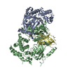
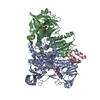
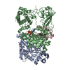
Mass: 71282.445 Da / Num. of mol.: 2 Source method: isolated from a genetically manipulated source Source: (gene. exp.) Homo sapiens (human) / Gene: UBLE1B, SAE2 / Plasmid: PET28B / Production host: Escherichia coli (E. coli) / Strain (production host): BL21DE3CP / References: UniProt: Q9UBT2
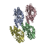
#3: Protein
Ubiquitin-likeproteinSMT3C / Ubiquitin-homology domain protein PIC1 / Ubiquitin-like protein UBL1 / Ubiquitin-related protein ...Ubiquitin-homology domain protein PIC1 / Ubiquitin-like protein UBL1 / Ubiquitin-related protein SUMO-1 / GAP modifying protein 1 / GMP1 / Sentrin / OK/SW-cl.43
Mass: 11149.545 Da / Num. of mol.: 2 Source method: isolated from a genetically manipulated source Source: (gene. exp.) Homo sapiens (human) / Gene: UBL1, SUMO-1, SMT3C, SMT3H3 / Plasmid: PET28B / Species (production host): Escherichia coli / Production host: Escherichia coli BL21(DE3) (bacteria) / Strain (production host): BL21DE3 / References: UniProt: P63165
In the structure databanks used in Yorodumi, some data are registered as the other names, "COVID-19 virus" and "2019-nCoV". Here are the details of the virus and the list of structure data.
Jan 31, 2019. EMDB accession codes are about to change! (news from PDBe EMDB page)
EMDB accession codes are about to change! (news from PDBe EMDB page)
The allocation of 4 digits for EMDB accession codes will soon come to an end. Whilst these codes will remain in use, new EMDB accession codes will include an additional digit and will expand incrementally as the available range of codes is exhausted. The current 4-digit format prefixed with “EMD-” (i.e. EMD-XXXX) will advance to a 5-digit format (i.e. EMD-XXXXX), and so on. It is currently estimated that the 4-digit codes will be depleted around Spring 2019, at which point the 5-digit format will come into force.
The EM Navigator/Yorodumi systems omit the EMD- prefix.
Related info.:Q: What is EMD? / ID/Accession-code notation in Yorodumi/EM Navigator
Yorodumi is a browser for structure data from EMDB, PDB, SASBDB, etc.
This page is also the successor to EM Navigator detail page, and also detail information page/front-end page for Omokage search.
The word "yorodu" (or yorozu) is an old Japanese word meaning "ten thousand". "mi" (miru) is to see.
Related info.:EMDB / PDB / SASBDB / Comparison of 3 databanks / Yorodumi Search / Aug 31, 2016. New EM Navigator & Yorodumi / Yorodumi Papers / Jmol/JSmol / Function and homology information / Changes in new EM Navigator and Yorodumi
 Movie
Movie Controller


 Open data
 Basic information
 Components
 Keywords
 Function and homology information
 Homo sapiens (human)
 X-RAY DIFFRACTION /
  Authors
 Citation
 Structure visualization
 Downloads & links
 Other downloads

 PDBj
PDBj


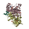
 Assembly
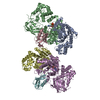
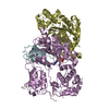


 Mass: 24.305 Da / Num. of mol.: 2 / Source method: obtained synthetically / Formula: Mg
 Mass: 65.409 Da / Num. of mol.: 2 / Source method: obtained synthetically / Formula: Zn
 Mass: 507.181 Da / Num. of mol.: 2 / Source method: obtained synthetically / Formula: C10H16N5O13P3 / Comment: ATP, energy-carrying molecule*YM
 Sample preparation
 / Beamline: 31-ID / Wavelength: 0.979 Å
 Processing