 Movie
Movie Controller

[English] 日本語
 Yorodumi
- PDB-2f5z: Crystal Structure of Human Dihydrolipoamide Dehydrogenase (E3) Co... -
+ Open data

- Basic information
| Entry | Database: PDB / ID: 2f5z | ||||||
|---|---|---|---|---|---|---|---|
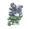
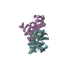
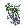
| Title | Crystal Structure of Human Dihydrolipoamide Dehydrogenase (E3) Complexed to the E3-Binding Domain of Human E3-Binding Protein | ||||||
 Components Components |
| ||||||
 Keywords Keywords | OXIDOREDUCTASE/PROTEIN BINDING / protein-protein complex / OXIDOREDUCTASE-PROTEIN BINDING COMPLEX | ||||||
| Function / homology |  Function and homology information Function and homology information: / acrosomal matrix / OGDH complex synthesizes succinyl-CoA from 2-OG / OADH complex synthesizes glutaryl-CoA from 2-OA / 2-oxoglutarate decarboxylation to succinyl-CoA / oxoadipate dehydrogenase complex / Glycine degradation / BCKDH synthesizes BCAA-CoA from KIC, KMVA, KIV / Loss-of-function mutations in DBT cause MSUD2 / Loss-of-function mutations in DLD cause MSUD3/DLDD ...: / acrosomal matrix / OGDH complex synthesizes succinyl-CoA from 2-OG / OADH complex synthesizes glutaryl-CoA from 2-OA / 2-oxoglutarate decarboxylation to succinyl-CoA / oxoadipate dehydrogenase complex / Glycine degradation / BCKDH synthesizes BCAA-CoA from KIC, KMVA, KIV / Loss-of-function mutations in DBT cause MSUD2 / Loss-of-function mutations in DLD cause MSUD3/DLDD / H139Hfs13* PPM1K causes a mild variant of MSUD / branched-chain alpha-keto acid decarboxylation to branched-chain acyl-CoA / branched-chain alpha-ketoacid dehydrogenase complex / PDH complex synthesizes acetyl-CoA from PYR / Branched-chain ketoacid dehydrogenase kinase deficiency / dihydrolipoyl dehydrogenase / dihydrolipoyl dehydrogenase (NADH) activity / dihydrolipoyllysine-residue acetyltransferase activity / pyruvate decarboxylation to acetyl-CoA / Regulation of pyruvate dehydrogenase (PDH) complex / oxoglutarate dehydrogenase complex / pyruvate dehydrogenase complex / Branched-chain amino acid catabolism / branched-chain amino acid catabolic process / 2-oxoglutarate metabolic process / motile cilium / Signaling by Retinoic Acid / sperm capacitation / mitochondrial electron transport, NADH to ubiquinone / gastrulation / Mitochondrial protein degradation / regulation of membrane potential / flavin adenine dinucleotide binding / mitochondrial matrix / mitochondrion / proteolysis / nucleoplasm / nucleus / plasma membrane Similarity search - Function | ||||||
| Biological species |  Homo sapiens (human) Homo sapiens (human) | ||||||
| Method |  X-RAY DIFFRACTION / SYNCHROTRON / MOLECULAR REPLACEMENT / Resolution: 2.18 Å X-RAY DIFFRACTION / SYNCHROTRON / MOLECULAR REPLACEMENT / Resolution: 2.18 Å | ||||||
 Authors Authors | Brautigam, C.A. / Chuang, J.L. / Wynn, R.M. / Tomchick, D.R. / Machius, M. / Chuang, D.T. | ||||||
 Citation Citation | Journal: Structure / Year: 2006 Title: Structural Insight into Interactions between Dihydrolipoamide Dehydrogenase (E3) and E3 Binding Protein of Human Pyruvate Dehydrogenase Complex. Authors: Brautigam, C.A. / Wynn, R.M. / Chuang, J.L. / Machius, M. / Tomchick, D.R. / Chuang, D.T. | ||||||
| History |
| ||||||
| Remark 300 | BIOMOLECULE: 1, 2, 3, 4, 5 THIS ENTRY CONTAINS THE CRYSTALLOGRAPHIC ASYMMETRIC UNIT WHICH CONSISTS ...BIOMOLECULE: 1, 2, 3, 4, 5 THIS ENTRY CONTAINS THE CRYSTALLOGRAPHIC ASYMMETRIC UNIT WHICH CONSISTS OF 15CHAIN(S). SEE REMARK 350 FOR INFORMATION ON GENERATING THE BIOLOGICAL MOLECULE(S). AUTHORS INDICATE THAT BIOMOLECULE 1 BEST REPRESENTS THE PHYSIOLOGICAL COMPLEX |
- Structure visualization
| Structure viewer | Molecule: MolmilJmol/JSmol |
|---|
- Downloads & links
-Download
| PDBx/mmCIF format | 2f5z.cif.gz | 978.3 KB | Display | PDBx/mmCIF format |
|---|---|---|---|---|
| PDB format | pdb2f5z.ent.gz | 807.8 KB | Display | PDB format |
| PDBx/mmJSON format | 2f5z.json.gz | Tree view | PDBx/mmJSON format | |
| Others |  Other downloads Other downloads |
-Validation report
| Arichive directory | https://data.pdbj.org/pub/pdb/validation_reports/f5/2f5zftp://data.pdbj.org/pub/pdb/validation_reports/f5/2f5z | HTTPS FTP |
|---|
-Related structure data
| Related structure data | 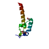 2f60C  1zmdS S: Starting model for refinement C: citing same article ( |
|---|---|
| Similar structure data |


-Links
 PDBj
PDBj


- Assembly
| Deposited unit | 
| ||||||||
|---|---|---|---|---|---|---|---|---|---|
| 1 | 
| ||||||||
| 2 | 
| ||||||||
| 3 | 
| ||||||||
| 4 | 
| ||||||||
| 5 | 
| ||||||||
| Unit cell |
|
-Components
| #1: Protein | Mass: 50208.406 Da / Num. of mol.: 10 / Fragment: Dihydrolipoyl dehydrogenase, residues 36-509 Source method: isolated from a genetically manipulated source Source: (gene. exp.) Homo sapiens (human) / Gene: DLD, GCSL, LAD, PHE3 / Plasmid: pTrcHisThE3 / Production host:  #2: Protein | Mass: 7150.151 Da / Num. of mol.: 5 / Fragment: E3-binding domain, residues 173-230 / Mutation: K120G,T176L Source method: isolated from a genetically manipulated source Source: (gene. exp.) Homo sapiens (human) / Gene: PDHX, PDX1 / Plasmid: pET-LTE3BD / Species (production host): Escherichia coli / Production host: #3: Chemical | ChemComp-SO4 /   Mass: 96.063 Da / Num. of mol.: 39 / Source method: obtained synthetically / Formula: SO4 Mass: 96.063 Da / Num. of mol.: 39 / Source method: obtained synthetically / Formula: SO4#4: Chemical | ChemComp-FAD /   Mass: 785.550 Da / Num. of mol.: 10 / Source method: obtained synthetically / Formula: C27H33N9O15P2 / Comment: FAD*YM Mass: 785.550 Da / Num. of mol.: 10 / Source method: obtained synthetically / Formula: C27H33N9O15P2 / Comment: FAD*YM#5: Water | ChemComp-HOH / |  Mass: 18.015 Da / Num. of mol.: 1776 / Source method: isolated from a natural source / Formula: H2O Mass: 18.015 Da / Num. of mol.: 1776 / Source method: isolated from a natural source / Formula: H2OHas protein modification | Y | |
|---|
-Experimental details
-Experiment
| Experiment | Method: X-RAY DIFFRACTION / Number of used crystals: 1 |
|---|
- Sample preparation
| Crystal | Density Matthews: 3.35 Å3/Da / Density % sol: 63.29 % |
|---|---|
| Crystal grow | Temperature: 293 K / Method: vapor diffusion, hanging drop / pH: 4.6 Details: 14% (w/v) PEG4000, 0.2 M ammonium sulfate, 0.1 M sodium acetate, 0.03 M spermidine, 8% (v/v) glycerol, pH 4.6, VAPOR DIFFUSION, HANGING DROP, temperature 293K |
-Data collection
| Diffraction | Mean temperature: 100 K |
|---|---|
| Diffraction source | Source: SYNCHROTRON / Site: APS  / Beamline: 19-ID / Wavelength: 0.979426 Å / Beamline: 19-ID / Wavelength: 0.979426 Å |
| Detector | Type: CUSTOM-MADE / Detector: CCD / Date: Jul 9, 2004 |
| Radiation | Monochromator: Si (111) / Protocol: SINGLE WAVELENGTH / Monochromatic (M) / Laue (L): M / Scattering type: x-ray |
| Radiation wavelength | Wavelength: 0.979426 Å / Relative weight: 1 |
| Reflection | Resolution: 2.18→37.4 Å / Num. all: 367237 / Num. obs: 367237 / % possible obs: 98.2 % / Observed criterion σ(I): -3 / Redundancy: 3.9 % / Biso Wilson estimate: 31.9 Å2 / Rsym value: 0.054 / Net I/σ(I): 21.9 |
| Reflection shell | Resolution: 2.18→2.26 Å / Redundancy: 2.5 % / Mean I/σ(I) obs: 1.9 / Num. unique all: 31492 / Rsym value: 0.401 / % possible all: 84.8 |
- Processing
| Software |
| |||||||||||||||||||||||||
|---|---|---|---|---|---|---|---|---|---|---|---|---|---|---|---|---|---|---|---|---|---|---|---|---|---|---|
| Refinement | Method to determine structure: MOLECULAR REPLACEMENT Starting model: PDB ENTRY 1ZMD without NADH Resolution: 2.18→37.4 Å / Isotropic thermal model: restrained / Cross valid method: THROUGHOUT / σ(F): 0 / Stereochemistry target values: Engh & Huber Details: Residues of E3BD with missing atoms were modeled as alanine due to missing density for the side chain
| |||||||||||||||||||||||||
| Displacement parameters | Biso mean: 42.3 Å2
| |||||||||||||||||||||||||
| Refine analyze |
| |||||||||||||||||||||||||
| Refinement step | Cycle: LAST / Resolution: 2.18→37.4 Å
| |||||||||||||||||||||||||
| Refine LS restraints |
| |||||||||||||||||||||||||
| LS refinement shell | Resolution: 2.18→2.32 Å / Rfactor Rfree error: 0.012
|
