Mass: 18.015 Da / Num. of mol.: 290 / Source method: isolated from a natural source / Formula: H2O
Has protein modification
Y
Sequence details
N-ACETYLATION IN THE STRUCTURE, NUMBERED AS RESIDUE ACE A 0 ERROR IN CDNA SEQUENCING GLN142 WRITTEN ...N-ACETYLATION IN THE STRUCTURE, NUMBERED AS RESIDUE ACE A 0 ERROR IN CDNA SEQUENCING GLN142 WRITTEN AS PRO IN DATABASE (UNDER CORRECTION)
-
Experimental details
-
Experiment
Experiment
Method: X-RAY DIFFRACTION / Number of used crystals: 2
-
Sample preparation
Crystal
Density Matthews: 3.6 Å3/Da / Density % sol: 66.2 %
Crystal grow
Method: vapor diffusion, hanging drop / pH: 7.5 Details: HEPES PH 7.5, 200MM CACL2, 14 TO 20% PEG 550, pH 7.5, vapor diffusion, hanging drop, VAPOR DIFFUSION, HANGING DROP
Crystal grow
*PLUS
Method: unknown / Details: Petitpas, I., (1998) J.Virol., 72, 7615.
Components of the solutions
*PLUS
ID
Conc.
Common name
Crystal-ID
Sol-ID
Chemical formula
1
10-20 mg/ml
protein
1
1
2
100mM
1
1
Ca2+
3
14-20 %
mPEG550
1
1
4
200mM
1
1
CaCl2
-
Data collection
Diffraction
ID
Mean temperature (K)
Crystal-ID
1
100
1
2
1
1,2
1
Diffraction source
Source
Site
Beamline
ID
Wavelength
SYNCHROTRON
EMBL/DESY, HAMBURG
X11
1
0.88
SYNCHROTRON
EMBL/DESY, HAMBURG
BW7B
2
0.88
Detector
Type
ID
Detector
Date
MARRESEARCH
1
IMAGE PLATE
Aug 15, 1998
2
Radiation
ID
Protocol
Monochromatic (M) / Laue (L)
Scattering type
Wavelength-ID
1
SINGLEWAVELENGTH
M
x-ray
1
2
SINGLEWAVELENGTH
M
x-ray
1
Radiation wavelength
Wavelength: 0.88 Å / Relative weight: 1
Reflection
Resolution: 1.95→42 Å / Num. obs: 48534 / % possible obs: 98.3 % / Redundancy: 7.1 % / Biso Wilson estimate: 28.11 Å2 / Rsym value: 7.2 / Net I/σ(I): 15.6
Reflection shell
Resolution: 1.95→1.98 Å / Redundancy: 3.8 % / Mean I/σ(I) obs: 5.1 / Rsym value: 25.2 / % possible all: 97.3
Reflection
*PLUS
Rmerge(I) obs: 0.072
Reflection shell
*PLUS
% possible obs: 97.3 % / Rmerge(I) obs: 0.252
-
Processing
Software
Name
Version
Classification
SHARP
phasing
X-PLOR
3.851
refinement
DENZO
datareduction
SCALEPACK
datascaling
Refinement
Method to determine structure: MIRAS / Resolution: 1.95→42 Å / Rfactor Rfree error: 0.0044 / Isotropic thermal model: RESTRAINED / Cross valid method: THROUGHOUT / σ(F): 0
Rfactor
Num. reflection
% reflection
Selection details
Rfree
0.22
2444
5 %
RANDOM
Rwork
0.188
-
-
-
obs
-
48467
98.3 %
-
Displacement parameters
Biso mean: 27.04 Å2
Refine analyze
Free
Obs
Luzzati coordinate error
0.252 Å
0.21 Å
Luzzati d res low
-
5 Å
Luzzati sigma a
0.209 Å
0.22 Å
Refinement step
Cycle: LAST / Resolution: 1.95→42 Å
Protein
Nucleic acid
Ligand
Solvent
Total
Num. atoms
3162
0
7
290
3459
Refine LS restraints
Refine-ID
Type
Dev ideal
Dev ideal target
X-RAY DIFFRACTION
x_bond_d
0.012
X-RAY DIFFRACTION
x_bond_d_na
X-RAY DIFFRACTION
x_bond_d_prot
X-RAY DIFFRACTION
x_angle_d
X-RAY DIFFRACTION
x_angle_d_na
X-RAY DIFFRACTION
x_angle_d_prot
X-RAY DIFFRACTION
x_angle_deg
1.629
X-RAY DIFFRACTION
x_angle_deg_na
X-RAY DIFFRACTION
x_angle_deg_prot
X-RAY DIFFRACTION
x_dihedral_angle_d
24.94
X-RAY DIFFRACTION
x_dihedral_angle_d_na
X-RAY DIFFRACTION
x_dihedral_angle_d_prot
X-RAY DIFFRACTION
x_improper_angle_d
1.425
X-RAY DIFFRACTION
x_improper_angle_d_na
X-RAY DIFFRACTION
x_improper_angle_d_prot
X-RAY DIFFRACTION
x_mcbond_it
1.367
1
X-RAY DIFFRACTION
x_mcangle_it
2.297
1
X-RAY DIFFRACTION
x_scbond_it
3.876
2
X-RAY DIFFRACTION
x_scangle_it
6.01
2
LS refinement shell
Resolution: 1.95→2.02 Å / Rfactor Rfree error: 0.016 / Total num. of bins used: 10
In the structure databanks used in Yorodumi, some data are registered as the other names, "COVID-19 virus" and "2019-nCoV". Here are the details of the virus and the list of structure data.
Jan 31, 2019. EMDB accession codes are about to change! (news from PDBe EMDB page)
EMDB accession codes are about to change! (news from PDBe EMDB page)
The allocation of 4 digits for EMDB accession codes will soon come to an end. Whilst these codes will remain in use, new EMDB accession codes will include an additional digit and will expand incrementally as the available range of codes is exhausted. The current 4-digit format prefixed with “EMD-” (i.e. EMD-XXXX) will advance to a 5-digit format (i.e. EMD-XXXXX), and so on. It is currently estimated that the 4-digit codes will be depleted around Spring 2019, at which point the 5-digit format will come into force.
The EM Navigator/Yorodumi systems omit the EMD- prefix.
Related info.:Q: What is EMD? / ID/Accession-code notation in Yorodumi/EM Navigator
Yorodumi is a browser for structure data from EMDB, PDB, SASBDB, etc.
This page is also the successor to EM Navigator detail page, and also detail information page/front-end page for Omokage search.
The word "yorodu" (or yorozu) is an old Japanese word meaning "ten thousand". "mi" (miru) is to see.
Related info.:EMDB / PDB / SASBDB / Comparison of 3 databanks / Yorodumi Search / Aug 31, 2016. New EM Navigator & Yorodumi / Yorodumi Papers / Jmol/JSmol / Function and homology information / Changes in new EM Navigator and Yorodumi
 Movie
Movie Controller

 Yorodumi
 Open data
 Basic information
 Components
 Keywords
 Function and homology information
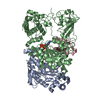
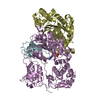
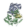
 Bovine rotavirus
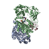
 X-RAY DIFFRACTION /
  Authors
 Citation
 Structure visualization
 Downloads & links
 Other downloads


 PDBj
PDBj


 Assembly
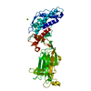

 Spodoptera frugiperda (fall armyworm) / References: UniProt: P04509
 Mass: 65.409 Da / Num. of mol.: 1 / Source method: obtained synthetically / Formula: Zn
 Mass: 35.453 Da / Num. of mol.: 1 / Source method: obtained synthetically / Formula: Cl
 Mass: 40.078 Da / Num. of mol.: 5 / Source method: obtained synthetically / Formula: Ca
 Mass: 18.015 Da / Num. of mol.: 290 / Source method: isolated from a natural source / Formula: H2O
 Sample preparation
 Processing