 Movie
Movie Controller


+ Open data

- Basic information
| Entry | Database: PDB / ID: 3kyc | ||||||
|---|---|---|---|---|---|---|---|
| Title | Human SUMO E1 complex with a SUMO1-AMP mimic | ||||||
 Components Components |
| ||||||
 Keywords Keywords | LIGASE / E1 / SUMO / UBIQUITIN / THIOESTER / ADENYLATION / INHIBITOR / ACYL-ADENYLATE INTERMEDIATE / Acetylation / Nucleus / Phosphoprotein / Ubl conjugation pathway / ATP-binding / Nucleotide-binding / Polymorphism / Cytoplasm / Isopeptide bond / Membrane | ||||||
| Function / homology |  Function and homology information Function and homology informationSUMO activating enzyme activity / SUMO activating enzyme complex / SUMOylation of nuclear envelope proteins / SUMO is proteolytically processed / Negative regulation of activity of TFAP2 (AP-2) family transcription factors / SUMO is conjugated to E1 (UBA2:SAE1) / ubiquitin activating enzyme activity / SUMO is transferred from E1 to E2 (UBE2I, UBC9) / negative regulation of transcription initiation by RNA polymerase II / negative regulation of action potential ...SUMO activating enzyme activity / SUMO activating enzyme complex / SUMOylation of nuclear envelope proteins / SUMO is proteolytically processed / Negative regulation of activity of TFAP2 (AP-2) family transcription factors / SUMO is conjugated to E1 (UBA2:SAE1) / ubiquitin activating enzyme activity / SUMO is transferred from E1 to E2 (UBE2I, UBC9) / negative regulation of transcription initiation by RNA polymerase II / negative regulation of action potential / nuclear stress granule / positive regulation of protein sumoylation / small protein activating enzyme binding / SUMO binding / SUMOylation of immune response proteins / SUMOylation of SUMOylation proteins / regulation of calcium ion transmembrane transport / SUMOylation of DNA methylation proteins / Maturation of nucleoprotein / XY body / ATP-dependent protein binding / SUMOylation of RNA binding proteins / Transferases; Acyltransferases; Aminoacyltransferases / regulation of cardiac muscle cell contraction / Postmitotic nuclear pore complex (NPC) reformation / Maturation of nucleoprotein / : / negative regulation of protein import into nucleus / roof of mouth development / Maturation of DENV proteins / SUMOylation of ubiquitinylation proteins / ubiquitin-like protein conjugating enzyme binding / ubiquitin-specific protease binding / cellular response to cadmium ion / SUMOylation of transcription factors / SUMOylation of DNA replication proteins / ubiquitin-like protein ligase binding / protein sumoylation / nuclear pore / potassium channel regulator activity / Regulation of IFNG signaling / transporter activator activity / postsynaptic cytosol / SUMOylation of DNA damage response and repair proteins / presynaptic cytosol / Transcriptional and post-translational regulation of MITF-M expression and activity / SUMOylation of transcription cofactors / SUMOylation of chromatin organization proteins / SUMOylation of intracellular receptors / regulation of protein stability / positive regulation of protein-containing complex assembly / PML body / PKR-mediated signaling / enzyme activator activity / Formation of Incision Complex in GG-NER / protein tag activity / positive regulation of proteasomal ubiquitin-dependent protein catabolic process / cellular response to heat / Recruitment and ATM-mediated phosphorylation of repair and signaling proteins at DNA double strand breaks / transferase activity / nuclear membrane / nuclear speck / nuclear body / protein stabilization / protein heterodimerization activity / negative regulation of DNA-templated transcription / DNA repair / ubiquitin protein ligase binding / nucleolus / glutamatergic synapse / magnesium ion binding / enzyme binding / negative regulation of transcription by RNA polymerase II / RNA binding / nucleoplasm / ATP binding / nucleus / plasma membrane / cytosol / cytoplasm Similarity search - Function | ||||||
| Biological species |  Homo sapiens (human) Homo sapiens (human) | ||||||
| Method |  X-RAY DIFFRACTION / SYNCHROTRON / MOLECULAR REPLACEMENT / molecular replacement / Resolution: 2.45 Å X-RAY DIFFRACTION / SYNCHROTRON / MOLECULAR REPLACEMENT / molecular replacement / Resolution: 2.45 Å | ||||||
 Authors Authors | Lima, C.D. | ||||||
 Citation Citation | Journal: Nature / Year: 2010 Title: Active site remodelling accompanies thioester bond formation in the SUMO E1. Authors: Olsen, S.K. / Capili, A.D. / Lu, X. / Tan, D.S. / Lima, C.D. | ||||||
| History |
|
- Structure visualization
| Structure viewer | Molecule: MolmilJmol/JSmol |
|---|
- Downloads & links
-Download
| PDBx/mmCIF format | 3kyc.cif.gz | 210.1 KB | Display | PDBx/mmCIF format |
|---|---|---|---|---|
| PDB format | pdb3kyc.ent.gz | 161.5 KB | Display | PDB format |
| PDBx/mmJSON format | 3kyc.json.gz | Tree view | PDBx/mmJSON format | |
| Others |  Other downloads Other downloads |
-Validation report
| Arichive directory | https://data.pdbj.org/pub/pdb/validation_reports/ky/3kycftp://data.pdbj.org/pub/pdb/validation_reports/ky/3kyc | HTTPS FTP |
|---|
-Related structure data
| Related structure data | 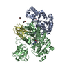 3kydC  1y8qS C: citing same article ( S: Starting model for refinement |
|---|---|
| Similar structure data |


-Links
 PDBj
PDBj


- Assembly
| Deposited unit | 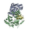
| ||||||||
|---|---|---|---|---|---|---|---|---|---|
| 1 |
| ||||||||
| Unit cell |
| ||||||||
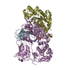
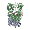
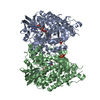
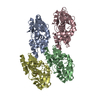
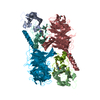
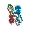
| Details | The biological unit is a heterotrimer. |
-Components
-SUMO-activating enzyme subunit ... , 2 types, 2 molecules AB
| #1: Protein | Mass: 38499.789 Da / Num. of mol.: 1 Source method: isolated from a genetically manipulated source Source: (gene. exp.) Homo sapiens (human) / Gene: AOS1, SAE1, SUA1, UBLE1A / Plasmid: pET-15b / Production host:  |
|---|---|
| #2: Protein | Mass: 73501.953 Da / Num. of mol.: 1 Source method: isolated from a genetically manipulated source Source: (gene. exp.) Homo sapiens (human) / Gene: HRIHFB2115, SAE2, UBA2, UBLE1B / Plasmid: pET28b / Production host: References: UniProt: Q9UBT2, Ligases; Forming carbon-nitrogen bonds; Acid-amino-acid ligases (peptide synthases) |
-Protein , 1 types, 1 molecules D
| #3: Protein | Mass: 11151.584 Da / Num. of mol.: 1 / Fragment: UNP residues 1-97 / Mutation: T95C Source method: isolated from a genetically manipulated source Source: (gene. exp.) Homo sapiens (human) / Gene: OK/SW-cl.43, SMT3C, SMT3H3, SUMO1, UBL1 / Plasmid: pTXB1 / Production host: |
|---|
-Non-polymers , 3 types, 255 molecules 


| #4: Chemical | ChemComp-ZN /  Mass: 65.409 Da / Num. of mol.: 1 / Source method: obtained synthetically / Formula: Zn Mass: 65.409 Da / Num. of mol.: 1 / Source method: obtained synthetically / Formula: ZnDetails: cysteylglycylglycyl 5'-(sulfamoylaminodeoxy)adenosine |
|---|---|
| #5: Chemical | ChemComp-JZU /  Mass: 345.335 Da / Num. of mol.: 1 / Source method: obtained synthetically / Formula: C10H15N7O5S Mass: 345.335 Da / Num. of mol.: 1 / Source method: obtained synthetically / Formula: C10H15N7O5S |
| #6: Water | ChemComp-HOH / Mass: 18.015 Da / Num. of mol.: 253 / Source method: isolated from a natural source / Formula: H2O |
-Details
| Has protein modification | Y |
|---|---|
| Nonpolymer details | LIGAND CYS-GLY-GLY-AMSN, WHERE AMSN HAS ID JZU IN THIS FILE, WAS LINKED TO SUMO1 BY INTEIN-MEDIATED ...LIGAND CYS-GLY-GLY-AMSN, WHERE AMSN HAS ID JZU IN THIS FILE, WAS LINKED TO SUMO1 BY INTEIN-MEDIATED LIGATION TO MAKE CHAIN D. |
| Sequence details | CHAIN D IS COMPOSED AS UB/UBI-CYS-GLY-GLY-AMSN, ADENYLATE MIMIC. |
-Experimental details
-Experiment
| Experiment | Method: X-RAY DIFFRACTION / Number of used crystals: 1 |
|---|
- Sample preparation
| Crystal | Density Matthews: 2.56 Å3/Da / Density % sol: 51.91 % |
|---|---|
| Crystal grow | Temperature: 291 K / Method: vapor diffusion, hanging drop / pH: 6.4 Details: 0.1 MES, 24% PEG 3350, 1.3 M AMMONIUM ACETATE, 10 mM ATP, 10 mM MgCl2, 10 mM TCEP, 2.5% PEG 400, pH 6.4, VAPOR DIFFUSION, HANGING DROP, temperature 291K |
-Data collection
| Diffraction | Mean temperature: 100 K |
|---|---|
| Diffraction source | Source: SYNCHROTRON / Site: APS  / Beamline: 24-ID-C / Wavelength: 0.979 Å / Beamline: 24-ID-C / Wavelength: 0.979 Å |
| Detector | Type: ADSC QUANTUM 315 / Detector: CCD / Date: Nov 26, 2007 |
| Radiation | Protocol: SINGLE WAVELENGTH / Monochromatic (M) / Laue (L): M / Scattering type: x-ray |
| Radiation wavelength | Wavelength: 0.979 Å / Relative weight: 1 |
| Reflection | Resolution: 2.45→25 Å / Num. obs: 47106 / % possible obs: 99.2 % / Observed criterion σ(I): 0 / Rmerge(I) obs: 0.052 / Net I/σ(I): 12.3 |
| Reflection shell | Resolution: 2.45→2.51 Å / Rmerge(I) obs: 0.356 / Mean I/σ(I) obs: 2.2 / % possible all: 98 |
-Phasing
| Phasing | Method: molecular replacement |
|---|
- Processing
| Software |
| |||||||||||||||||||||||||||||||||||||||||||||||||||||||||||||||||
|---|---|---|---|---|---|---|---|---|---|---|---|---|---|---|---|---|---|---|---|---|---|---|---|---|---|---|---|---|---|---|---|---|---|---|---|---|---|---|---|---|---|---|---|---|---|---|---|---|---|---|---|---|---|---|---|---|---|---|---|---|---|---|---|---|---|---|
| Refinement | Method to determine structure: MOLECULAR REPLACEMENT Starting model: PDB ENTRY 1Y8Q Resolution: 2.45→25 Å / Cor.coef. Fo:Fc: 0.962 / Cor.coef. Fo:Fc free: 0.93 / Occupancy max: 1 / Occupancy min: 1 / SU B: 5.958 / SU ML: 0.133 / Cross valid method: THROUGHOUT / σ(F): 0 / ESU R: 0.355 / ESU R Free: 0.259 / Stereochemistry target values: MAXIMUM LIKELIHOOD / Details: U VALUES : REFINED INDIVIDUALLY
| |||||||||||||||||||||||||||||||||||||||||||||||||||||||||||||||||
| Solvent computation | Ion probe radii: 1 Å / Shrinkage radii: 0.8 Å / VDW probe radii: 1 Å / Solvent model: BABINET MODEL WITH MASK | |||||||||||||||||||||||||||||||||||||||||||||||||||||||||||||||||
| Displacement parameters | Biso max: 149.48 Å2 / Biso mean: 71.486 Å2 / Biso min: 26.17 Å2
| |||||||||||||||||||||||||||||||||||||||||||||||||||||||||||||||||
| Refinement step | Cycle: LAST / Resolution: 2.45→25 Å
| |||||||||||||||||||||||||||||||||||||||||||||||||||||||||||||||||
| Refine LS restraints |
| |||||||||||||||||||||||||||||||||||||||||||||||||||||||||||||||||
| LS refinement shell | Resolution: 2.45→2.513 Å / Total num. of bins used: 20
|
