 Movie
Movie Controller

[English] 日本語
 Yorodumi
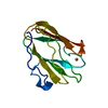
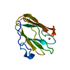
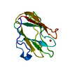
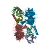
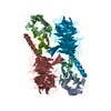
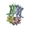
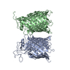
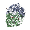
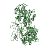
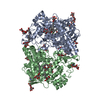
- PDB-2j55: X-ray reduced Paraccocus denitrificans methylamine dehydrogenase ... -
+ Open data

- Basic information
| Entry | Database: PDB / ID: 2j55 | ||||||
|---|---|---|---|---|---|---|---|
| Title | X-ray reduced Paraccocus denitrificans methylamine dehydrogenase O- quinone in complex with amicyanin. | ||||||
 Components Components |
| ||||||
 Keywords Keywords | OXIDOREDUCTASE / TRANSPORT / PERIPLASMIC / METAL-BINDING / ELECTRON TRANSPORT / SINGLE CRYSTAL MICROSPECTROPHOTOMETRY | ||||||
| Function / homology |  Function and homology information Function and homology informationmethylamine dehydrogenase (amicyanin) / methylamine dehydrogenase (amicyanin) activity / methylamine metabolic process / aliphatic amine dehydrogenase activity / amine metabolic process / outer membrane-bounded periplasmic space / electron transfer activity / periplasmic space / copper ion binding Similarity search - Function | ||||||
| Biological species |  PARACOCCUS DENITRIFICANS (bacteria) PARACOCCUS DENITRIFICANS (bacteria) | ||||||
| Method |  X-RAY DIFFRACTION / SYNCHROTRON / MOLECULAR REPLACEMENT / Resolution: 2.15 Å X-RAY DIFFRACTION / SYNCHROTRON / MOLECULAR REPLACEMENT / Resolution: 2.15 Å | ||||||
 Authors Authors | Pearson, A.R. / Pahl, R. / Davidson, V.L. / Wilmot, C.M. | ||||||
 Citation Citation | Journal: J.Synchrotron Radiat. / Year: 2007 Title: Tracking X-Ray-Derived Redox Changes in Crystals of a Methylamine Dehydrogenase/Amicyanin Complex Using Single-Crystal Uv/Vis Microspectrophotometry. Authors: Pearson, A.R. / Pahl, R. / Kovaleva, E.G. / Davidson, V.L. / Wilmot, C.M. #1: Journal: To be PublishedTitle: Crystallographic Structures of Methylamine Dehydrogenase Catalytic Intermediates from Paraccocus Denitrificans Authors: De La Mora-Rey, T. / Pearson, A.R. / Hoeffner, E. / Watts, K.T. / Yucel, N. / Davidson, V.L. / Wilmot, C.M. | ||||||
| History |
| ||||||
| Remark 650 | HELIX DETERMINATION METHOD: AUTHOR PROVIDED. | ||||||
| Remark 700 | SHEET THE SHEET STRUCTURE OF THIS MOLECULE IS BIFURCATED. IN ORDER TO REPRESENT THIS FEATURE IN ... SHEET THE SHEET STRUCTURE OF THIS MOLECULE IS BIFURCATED. IN ORDER TO REPRESENT THIS FEATURE IN THE SHEET RECORDS BELOW, TWO SHEETS ARE DEFINED. |
- Structure visualization
| Structure viewer | Molecule: MolmilJmol/JSmol |
|---|
- Downloads & links
-Download
| PDBx/mmCIF format | 2j55.cif.gz | 267.2 KB | Display | PDBx/mmCIF format |
|---|---|---|---|---|
| PDB format | pdb2j55.ent.gz | 214.5 KB | Display | PDB format |
| PDBx/mmJSON format | 2j55.json.gz | Tree view | PDBx/mmJSON format | |
| Others |  Other downloads Other downloads |
-Validation report
| Arichive directory | https://data.pdbj.org/pub/pdb/validation_reports/j5/2j55ftp://data.pdbj.org/pub/pdb/validation_reports/j5/2j55 | HTTPS FTP |
|---|
-Related structure data


-Links
 PDBj
PDBj

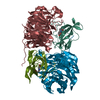
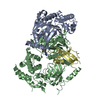
- Assembly
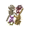
| Deposited unit | 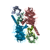
| ||||||||
|---|---|---|---|---|---|---|---|---|---|
| 1 |
| ||||||||
| Unit cell |
|
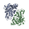
-Components
-Protein / METHYLAMINE DEHYDROGENASE ... / Antibody , 3 types, 6 molecules ABHJLM
| #1: Protein | Mass: 11505.171 Da / Num. of mol.: 2 / Source method: isolated from a natural source Details: AMICYANIN IS THE OBLIGATE ELECTRON TRANSFER PARTNER OF METHYLAMINE DEHYDROGENASE. Source: (natural) PARACOCCUS DENITRIFICANS (bacteria) / References: UniProt: P22364#2: Protein | Mass: 42449.277 Da / Num. of mol.: 2 / Source method: isolated from a natural source / Source: (natural) PARACOCCUS DENITRIFICANS (bacteria) / References: UniProt: P29894, EC: 1.4.99.3#3: Antibody | Mass: 14210.696 Da / Num. of mol.: 2 / Source method: isolated from a natural source / Source: (natural) PARACOCCUS DENITRIFICANS (bacteria) / References: UniProt: P22619, EC: 1.4.99.3 |
|---|
-Non-polymers , 3 types, 916 molecules 


| #4: Chemical |  Mass: 63.546 Da / Num. of mol.: 2 / Source method: obtained synthetically / Formula: Cu Mass: 63.546 Da / Num. of mol.: 2 / Source method: obtained synthetically / Formula: Cu#5: Chemical | ChemComp-GOL /  Mass: 92.094 Da / Num. of mol.: 4 / Source method: obtained synthetically / Formula: C3H8O3 Mass: 92.094 Da / Num. of mol.: 4 / Source method: obtained synthetically / Formula: C3H8O3#6: Water | ChemComp-HOH / | Mass: 18.015 Da / Num. of mol.: 910 / Source method: isolated from a natural source / Formula: H2O |
|---|
-Details
| Has protein modification | Y |
|---|---|
| Sequence details | AT PRESENT, THE SEQUENCE DATABASES INDICATE THAT RESIDUE 312 OF THE HEAVY CHAIN IS LEU AND RESIDUE ...AT PRESENT, THE SEQUENCE DATABASES INDICATE THAT RESIDUE 312 OF THE HEAVY CHAIN IS LEU AND RESIDUE 313 IS LEU. THE AUTHORS WHO DEPOSITED 2MTA FOUND THAT THEY MISREAD THE GELS AND THAT RESIDUES 312 AND 313 SHOULD BE PHE AND VAL, RESPECTIVE |
-Experimental details
-Experiment
| Experiment | Method: X-RAY DIFFRACTION / Number of used crystals: 5 |
|---|
- Sample preparation
| Crystal | Density Matthews: 3.32 Å3/Da / Density % sol: 62.95 % |
|---|---|
| Crystal grow | pH: 5.6 / Details: pH 5.60 |
-Data collection
| Diffraction | Mean temperature: 100 K |
|---|---|
| Diffraction source | Source: SYNCHROTRON / Site: APS  / Beamline: 14-BM-C / Wavelength: 0.9 / Beamline: 14-BM-C / Wavelength: 0.9 |
| Detector | Type: ADSC CCD / Detector: CCD |
| Radiation | Protocol: SINGLE WAVELENGTH / Monochromatic (M) / Laue (L): M / Scattering type: x-ray |
| Radiation wavelength | Wavelength: 0.9 Å / Relative weight: 1 |
| Reflection | Resolution: 2.15→21.7 Å / Num. obs: 96429 / % possible obs: 99 % / Observed criterion σ(I): 2 / Redundancy: 7.8 % / Biso Wilson estimate: 59.8 Å2 / Rmerge(I) obs: 0.11 / Net I/σ(I): 10.3 |
| Reflection shell | Resolution: 2.15→2.21 Å / Redundancy: 7.9 % / Rmerge(I) obs: 0.43 / Mean I/σ(I) obs: 5.2 / % possible all: 99.9 |
- Processing
| Software |
| ||||||||||||||||||||||||||||||||||||||||||||||||||||||||||||||||||||||||||||||||||||||||||||||||||||||||||||||||||||||||||||||||||||||||||||||||||||||||||||||||||||||||||||||||||||||
|---|---|---|---|---|---|---|---|---|---|---|---|---|---|---|---|---|---|---|---|---|---|---|---|---|---|---|---|---|---|---|---|---|---|---|---|---|---|---|---|---|---|---|---|---|---|---|---|---|---|---|---|---|---|---|---|---|---|---|---|---|---|---|---|---|---|---|---|---|---|---|---|---|---|---|---|---|---|---|---|---|---|---|---|---|---|---|---|---|---|---|---|---|---|---|---|---|---|---|---|---|---|---|---|---|---|---|---|---|---|---|---|---|---|---|---|---|---|---|---|---|---|---|---|---|---|---|---|---|---|---|---|---|---|---|---|---|---|---|---|---|---|---|---|---|---|---|---|---|---|---|---|---|---|---|---|---|---|---|---|---|---|---|---|---|---|---|---|---|---|---|---|---|---|---|---|---|---|---|---|---|---|---|---|
| Refinement | Method to determine structure: MOLECULAR REPLACEMENT Starting model: PRE-REDUCTION O-QUINONE FORM Resolution: 2.15→21.7 Å / Cor.coef. Fo:Fc: 0.958 / Cor.coef. Fo:Fc free: 0.932 / SU B: 9.369 / SU ML: 0.13 / Cross valid method: THROUGHOUT / ESU R: 0.181 / ESU R Free: 0.177 Stereochemistry target values: MAXIMUM LIKELIHOODWITH PHASES Details: HYDROGENS HAVE BEEN ADDED IN THE RIDING POSITIONS.
| ||||||||||||||||||||||||||||||||||||||||||||||||||||||||||||||||||||||||||||||||||||||||||||||||||||||||||||||||||||||||||||||||||||||||||||||||||||||||||||||||||||||||||||||||||||||
| Solvent computation | Ion probe radii: 0.8 Å / Shrinkage radii: 0.8 Å / VDW probe radii: 1.2 Å / Solvent model: MASK | ||||||||||||||||||||||||||||||||||||||||||||||||||||||||||||||||||||||||||||||||||||||||||||||||||||||||||||||||||||||||||||||||||||||||||||||||||||||||||||||||||||||||||||||||||||||
| Displacement parameters | Biso mean: 59.77 Å2
| ||||||||||||||||||||||||||||||||||||||||||||||||||||||||||||||||||||||||||||||||||||||||||||||||||||||||||||||||||||||||||||||||||||||||||||||||||||||||||||||||||||||||||||||||||||||
| Refinement step | Cycle: LAST / Resolution: 2.15→21.7 Å
| ||||||||||||||||||||||||||||||||||||||||||||||||||||||||||||||||||||||||||||||||||||||||||||||||||||||||||||||||||||||||||||||||||||||||||||||||||||||||||||||||||||||||||||||||||||||
| Refine LS restraints |
|