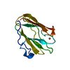
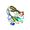
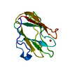
SHEET THE SHEET STRUCTURE OF THIS MOLECULE IS BIFURCATED. IN ORDER TO REPRESENT THIS FEATURE IN ... SHEET THE SHEET STRUCTURE OF THIS MOLECULE IS BIFURCATED. IN ORDER TO REPRESENT THIS FEATURE IN THE SHEET RECORDS BELOW, TWO SHEETS ARE DEFINED.
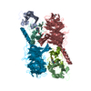
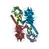
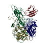
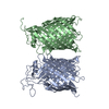
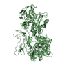
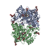
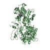
Mass: 11505.171 Da / Num. of mol.: 2 / Source method: isolated from a natural source Details: AMICYANIN IS THE OBLIGATE ELECTRON TRANSFER PARTNER OF METHYLAMINE DEHYDROGENASE. Source: (natural) PARACOCCUS DENITRIFICANS (bacteria) / References: UniProt: P22364
#2: Protein
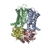
METHYLAMINEDEHYDROGENASEHEAVYCHAIN / METHYLAMINE DEHYDROGENASE ALPHA CHAIN / MADH
Mass: 42449.277 Da / Num. of mol.: 2 / Source method: isolated from a natural source / Source: (natural) PARACOCCUS DENITRIFICANS (bacteria) / References: UniProt: P29894, EC: 1.4.99.3
#3: Antibody
METHYLAMINEDEHYDROGENASELIGHTCHAIN / METHYLAMINE DEHYDROGENASE BETA CHAIN / MADH
Mass: 14209.712 Da / Num. of mol.: 2 / Source method: isolated from a natural source / Source: (natural) PARACOCCUS DENITRIFICANS (bacteria) / References: UniProt: P22619, EC: 1.4.99.3
Mass: 18.015 Da / Num. of mol.: 1033 / Source method: isolated from a natural source / Formula: H2O
-
Details
Has protein modification
Y
Sequence details
AT PRESENT, THE SEQUENCE DATABASES INDICATE THAT RESIDUE 312 OF THE HEAVY CHAIN IS LEU AND RESIDUE ...AT PRESENT, THE SEQUENCE DATABASES INDICATE THAT RESIDUE 312 OF THE HEAVY CHAIN IS LEU AND RESIDUE 313 IS LEU. THE AUTHORS WHO DEPOSITED 2MTA FOUND THAT THEY MISREAD THE GELS AND THAT RESIDUES 312 AND 313 SHOULD BE PHE AND VAL, RESPECTIVELY. IN THIS ENTRY THE SEQUENCE ERRORS HAVE BEEN CORRECTED.
-
Experimental details
-
Experiment
Experiment
Method: X-RAY DIFFRACTION / Number of used crystals: 7
-
Sample preparation
Crystal
Density Matthews: 3.31 Å3/Da / Density % sol: 62.92 %
Protocol: SINGLE WAVELENGTH / Monochromatic (M) / Laue (L): M / Scattering type: x-ray
Radiation wavelength
Wavelength: 0.9 Å / Relative weight: 1
Reflection
Resolution: 2.1→43.5 Å / Num. obs: 101798 / % possible obs: 97.4 % / Observed criterion σ(I): 2 / Redundancy: 9.7 % / Biso Wilson estimate: 29.3 Å2 / Rmerge(I) obs: 0.13 / Net I/σ(I): 22
Reflection shell
Resolution: 2.1→2.18 Å / Redundancy: 9.7 % / Rmerge(I) obs: 0.5 / Mean I/σ(I) obs: 7.8 / % possible all: 100
-
Processing
Software
Name
Version
Classification
REFMAC
5.2.0005
refinement
HKL-2000
datareduction
HKL-2000
datascaling
Refinement
Method to determine structure: OTHER / Resolution: 2.1→43.48 Å / Cor.coef. Fo:Fc: 0.96 / Cor.coef. Fo:Fc free: 0.943 / SU B: 6.525 / SU ML: 0.092 / Cross valid method: THROUGHOUT / ESU R: 0.152 / ESU R Free: 0.143 / Stereochemistry target values: MAXIMUM LIKELIHOOD / Details: HYDROGENS HAVE BEEN ADDED IN THE RIDING POSITIONS.
Rfactor
Num. reflection
% reflection
Selection details
Rfree
0.207
5353
5 %
RANDOM
Rwork
0.169
-
-
-
obs
0.171
101798
97.4 %
-
Solvent computation
Ion probe radii: 0.8 Å / Shrinkage radii: 0.8 Å / VDW probe radii: 1.2 Å / Solvent model: BABINET MODEL WITH MASK
 Movie
Movie Controller

 Yorodumi
 Open data
 Basic information
 Components
 Keywords
 Function and homology information
 PARACOCCUS DENITRIFICANS (bacteria)
 X-RAY DIFFRACTION /
  Authors
 Citation
 Structure visualization
 Downloads & links
 Other downloads


 PDBj
PDBj

 Assembly


 Mass: 63.546 Da / Num. of mol.: 2 / Source method: obtained synthetically / Formula: Cu
 Mass: 92.094 Da / Num. of mol.: 1 / Source method: obtained synthetically / Formula: C3H8O3
 Mass: 22.990 Da / Num. of mol.: 2 / Source method: obtained synthetically / Formula: Na
 Sample preparation
 / Beamline: 14-BM-C / Wavelength: 0.9
  Processing