 Movie
Movie Controller

[English] 日本語
 Yorodumi
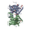
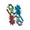
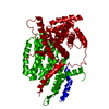
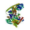
- PDB-1mda: CRYSTAL STRUCTURE OF AN ELECTRON-TRANSFER COMPLEX BETWEEN METHYLA... -
+ Open data

- Basic information
| Entry | Database: PDB / ID: 1mda | ||||||
|---|---|---|---|---|---|---|---|
| Title | CRYSTAL STRUCTURE OF AN ELECTRON-TRANSFER COMPLEX BETWEEN METHYLAMINE DEHYDROGENASE AND AMICYANIN | ||||||
 Components Components |
| ||||||
 Keywords Keywords | ELECTRON TRANSPORT | ||||||
| Function / homology |  Function and homology information Function and homology information | ||||||
| Biological species |  Paracoccus denitrificans (bacteria) Paracoccus denitrificans (bacteria) | ||||||
| Method |  X-RAY DIFFRACTION / Resolution: 2.5 Å X-RAY DIFFRACTION / Resolution: 2.5 Å | ||||||
 Authors Authors | Chen, L. / Durley, R. / Mathews, F.S. | ||||||
 Citation Citation | Journal: Biochemistry / Year: 1992 Title: Crystal structure of an electron-transfer complex between methylamine dehydrogenase and amicyanin. Authors: Chen, L. / Durley, R. / Poliks, B.J. / Hamada, K. / Chen, Z. / Mathews, F.S. / Davidson, V.L. / Satow, Y. / Huizinga, E. / Vellieux, F.M. / Hol, W.G.J. #1: Journal: Proteins / Year: 1992Title: Three-Dimensional Structure of the Quinoprotein Methylamine Dehydrogenase from Paracoccus Denitrificans Determined by Molecular Replacement at 2.8 Angstroms Resolution Authors: Chen, L. / Mathews, F.S. / Davidson, V.L. / Huizinga, E.G. / Vellieux, F.M.D. / Hol, W.G.J. #2: Journal: Science / Year: 1991Title: A New Cofactor in a Prokaryotic Enzyme: Tryptophan Tryptophylquinone as the Redox Prosthetic Group in Methylamine Dehydrogenase Authors: Mcintire, W.S. / Wemmer, D.E. / Chistoserdov, A. / Lidstrom, M.E. #3: Journal: Embo J. / Year: 1989Title: Structure of Quinoprotein Methylamine Dehydrogenase at 2.25 Angstroms Resolution Authors: Vellieux, F.M.D. / Huitema, F. / Groendijk, H. / Kalk, K.H. / Frank Jzn., J. / Jongejan, J.A. / Duine, J.A. / Petratos, K. / Drenth, J. / Hol, W.G.J. #4: Journal: Acta Crystallogr.,Sect.B / Year: 1990Title: Structure Determination of Quinoprotein Methylamine Dehydrogenase from Thiobacillus Versutus Authors: Vellieux, F.M.D. / Kalk, K.H. / Drenth, J. / Hol, W.G. | ||||||
| History |
|
- Structure visualization
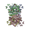
| Structure viewer | Molecule: MolmilJmol/JSmol |
|---|
- Downloads & links
-Download
| PDBx/mmCIF format | 1mda.cif.gz | 214.1 KB | Display | PDBx/mmCIF format |
|---|---|---|---|---|
| PDB format | pdb1mda.ent.gz | 167.8 KB | Display | PDB format |
| PDBx/mmJSON format | 1mda.json.gz | Tree view | PDBx/mmJSON format | |
| Others |  Other downloads Other downloads |
-Validation report
| Arichive directory | https://data.pdbj.org/pub/pdb/validation_reports/md/1mdaftp://data.pdbj.org/pub/pdb/validation_reports/md/1mda | HTTPS FTP |
|---|
-Related structure data
| Similar structure data |
|---|


-Links
 PDBj
PDBj- Assembly
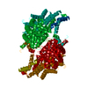
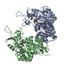
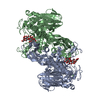
| Deposited unit | 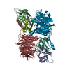
| ||||||||
|---|---|---|---|---|---|---|---|---|---|
| 1 |
| ||||||||
| Unit cell |
| ||||||||
| Atom site foot note | 1: CIS PROLINE - PRO A 6 / 2: CIS PROLINE - PRO B 6 | ||||||||
| Noncrystallographic symmetry (NCS) | NCS oper: (Code: given Matrix: (0.3439, -0.7653, -0.5441), Vector: Details | THE TRANSFORMATION PRESENTED ON *MTRIX* RECORDS BELOW WILL YIELD APPROXIMATE COORDINATES FOR CHAIN *H* AND *L* WHEN APPLIED TO CHAIN *J* AND *M*, RESPECTIVELY. | |
-Components
| #1: Protein | Mass: 36866.598 Da / Num. of mol.: 2 Source method: isolated from a genetically manipulated source Source: (gene. exp.) Paracoccus denitrificans (bacteria) / References: EC: 1.4.99.3#2: Protein | Mass: 13080.426 Da / Num. of mol.: 2 Source method: isolated from a genetically manipulated source Source: (gene. exp.) Paracoccus denitrificans (bacteria) / References: PIR: A44544, EC: 1.4.99.3#3: Protein | Mass: 11260.903 Da / Num. of mol.: 2 Source method: isolated from a genetically manipulated source Source: (gene. exp.) Paracoccus denitrificans (bacteria) / References: UniProt: P22364#4: Chemical | ChemComp-CU /   Mass: 63.546 Da / Num. of mol.: 2 / Source method: obtained synthetically / Formula: Cu Mass: 63.546 Da / Num. of mol.: 2 / Source method: obtained synthetically / Formula: CuCompound details | THE REDOX CENTERS OF MADH ARE LOCATED ON EACH L SUBUNIT. EACH IS COMPOSED OF THE SIDE CHAINS OF TWO ...THE REDOX CENTERS OF MADH ARE LOCATED ON EACH L SUBUNIT. EACH IS COMPOSED OF THE SIDE CHAINS OF TWO AMINO ACIDS ON THE L SUBUNIT, BOTH ARE TRYPTOPHAN | Sequence details | THIS IS AN X-RAY DETERMINED SEQUENCE WHICH WAS ESTABLISHED ON THE BASIS OF THE ELECTRON DENSITY DUE ...THIS IS AN X-RAY DETERMINED | |
|---|
-Experimental details
-Experiment
| Experiment | Method: X-RAY DIFFRACTION |
|---|
- Sample preparation
| Crystal | Density Matthews: 3.93 Å3/Da / Density % sol: 68.67 % | |||||||||||||||
|---|---|---|---|---|---|---|---|---|---|---|---|---|---|---|---|---|
| Crystal grow | *PLUS pH: 6.5 / Method: vapor diffusion, hanging drop | |||||||||||||||
| Components of the solutions | *PLUS
|
-Data collection
| Radiation | Scattering type: x-ray |
|---|---|
| Radiation wavelength | Relative weight: 1 |
| Reflection | *PLUS Highest resolution: 2.5 Å / Rmerge(I) obs: 0.084 |
- Processing
| Software |
| |||||||||||||||||||||||||||||||||||||||||||||||||||||||||||||||
|---|---|---|---|---|---|---|---|---|---|---|---|---|---|---|---|---|---|---|---|---|---|---|---|---|---|---|---|---|---|---|---|---|---|---|---|---|---|---|---|---|---|---|---|---|---|---|---|---|---|---|---|---|---|---|---|---|---|---|---|---|---|---|---|---|
| Refinement | Rfactor obs: 0.285 / Highest resolution: 2.5 Å | |||||||||||||||||||||||||||||||||||||||||||||||||||||||||||||||
| Refinement step | Cycle: LAST / Highest resolution: 2.5 Å
| |||||||||||||||||||||||||||||||||||||||||||||||||||||||||||||||
| Refine LS restraints |
| |||||||||||||||||||||||||||||||||||||||||||||||||||||||||||||||
| Refinement | *PLUS Highest resolution: 2.5 Å / Rfactor obs: 0.285 | |||||||||||||||||||||||||||||||||||||||||||||||||||||||||||||||
| Solvent computation | *PLUS | |||||||||||||||||||||||||||||||||||||||||||||||||||||||||||||||
| Displacement parameters | *PLUS Biso mean: 45 Å2 | |||||||||||||||||||||||||||||||||||||||||||||||||||||||||||||||
| Refine LS restraints | *PLUS
|
