Mass: 18.015 Da / Num. of mol.: 78 / Source method: isolated from a natural source / Formula: H2O
Has protein modification
Y
Nonpolymer details
2-MERCAPTOETHANOL (BME): COVALENTLY ATTACHED TO CYS A297
Sequence details
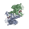
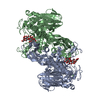
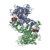
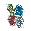
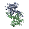
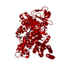
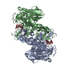
TRUNCATED CEP3P RESIDUES 48-608 THE TRUNCATED PROTEIN IS NUMBERED ACCORDING TO THE FULL LENGTH ...TRUNCATED CEP3P RESIDUES 48-608 THE TRUNCATED PROTEIN IS NUMBERED ACCORDING TO THE FULL LENGTH CEP3P SEQUENCE. DISULPHIDE LINK IS PRESENT BETWEEN A99 AND A215. DISORDERED REGIONS (N-TERMINUS-A48, A314- A335, A570-A587) WERE OMITTED FROM THE STRUCTURE. DISORDERED SIDE CHAINS OF UNDETERMINED ORIENTATION WERE GIVEN ZERO OCCUPANCY (65 ATOMS IN TOTAL)
-
Experimental details
-
Experiment
Experiment
Method: X-RAY DIFFRACTION / Number of used crystals: 1
-
Sample preparation
Crystal
Density Matthews: 3.29 Å3/Da / Density % sol: 59.8 % Description: SAD DATA COLLECTED FROM SELENOMETHIONINE- SUBSTITUTED PROTEIN
In the structure databanks used in Yorodumi, some data are registered as the other names, "COVID-19 virus" and "2019-nCoV". Here are the details of the virus and the list of structure data.
Jan 31, 2019. EMDB accession codes are about to change! (news from PDBe EMDB page)
EMDB accession codes are about to change! (news from PDBe EMDB page)
The allocation of 4 digits for EMDB accession codes will soon come to an end. Whilst these codes will remain in use, new EMDB accession codes will include an additional digit and will expand incrementally as the available range of codes is exhausted. The current 4-digit format prefixed with “EMD-” (i.e. EMD-XXXX) will advance to a 5-digit format (i.e. EMD-XXXXX), and so on. It is currently estimated that the 4-digit codes will be depleted around Spring 2019, at which point the 5-digit format will come into force.
The EM Navigator/Yorodumi systems omit the EMD- prefix.
Related info.:Q: What is EMD? / ID/Accession-code notation in Yorodumi/EM Navigator
Yorodumi is a browser for structure data from EMDB, PDB, SASBDB, etc.
This page is also the successor to EM Navigator detail page, and also detail information page/front-end page for Omokage search.
The word "yorodu" (or yorozu) is an old Japanese word meaning "ten thousand". "mi" (miru) is to see.
Related info.:EMDB / PDB / SASBDB / Comparison of 3 databanks / Yorodumi Search / Aug 31, 2016. New EM Navigator & Yorodumi / Yorodumi Papers / Jmol/JSmol / Function and homology information / Changes in new EM Navigator and Yorodumi
 Movie
Movie Controller

 Yorodumi
 Open data
 Basic information
 Components
 Keywords
 Function and homology information
 X-RAY DIFFRACTION /
  Authors
 Citation
 Structure visualization
 Downloads & links
 Other downloads
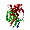


 PDBj
PDBj


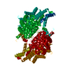
 Assembly
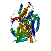


 Mass: 78.133 Da / Num. of mol.: 1 / Source method: obtained synthetically / Formula: C2H6OS
 Mass: 136.989 Da / Num. of mol.: 1 / Source method: obtained synthetically / Formula: C2H6AsO2
 Mass: 18.015 Da / Num. of mol.: 78 / Source method: isolated from a natural source / Formula: H2O
 Sample preparation
 / Beamline: ID29 / Wavelength: 1
  Processing