 Movie
Movie Controller

[English] 日本語
 Yorodumi
- PDB-1mg2: MUTATION OF ALPHA PHE55 OF METHYLAMINE DEHYDROGENASE ALTERS THE R... -
+ Open data

- Basic information
| Entry | Database: PDB / ID: 1mg2 | |||||||||
|---|---|---|---|---|---|---|---|---|---|---|
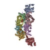
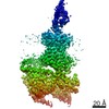
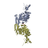
| Title | MUTATION OF ALPHA PHE55 OF METHYLAMINE DEHYDROGENASE ALTERS THE REORGANIZATION ENERGY AND ELECTRONIC COUPLING FOR ITS ELECTRON TRANSFER REACTION WITH AMICYANIN | |||||||||
 Components Components |
| |||||||||
 Keywords Keywords | OXIDOREDUCTASE / electron transfer / methylamine dehydrogenase / cytochrome / blue copper protein / active site mutant | |||||||||
| Function / homology |  Function and homology information Function and homology informationmicrotubule-dependent intracellular transport of viral material towards cell periphery / methylamine dehydrogenase (amicyanin) / methylamine dehydrogenase (amicyanin) activity / methanol metabolic process / methylamine metabolic process / aliphatic amine dehydrogenase activity / amine metabolic process / host cell / outer membrane-bounded periplasmic space / host cell endosome ...microtubule-dependent intracellular transport of viral material towards cell periphery / methylamine dehydrogenase (amicyanin) / methylamine dehydrogenase (amicyanin) activity / methanol metabolic process / methylamine metabolic process / aliphatic amine dehydrogenase activity / amine metabolic process / host cell / outer membrane-bounded periplasmic space / host cell endosome / periplasmic space / electron transfer activity / iron ion binding / copper ion binding / heme binding / virion membrane Similarity search - Function | |||||||||
| Biological species |  Paracoccus denitrificans (bacteria) Paracoccus denitrificans (bacteria) | |||||||||
| Method |  X-RAY DIFFRACTION / MOLECULAR REPLACEMENT / Resolution: 2.25 Å X-RAY DIFFRACTION / MOLECULAR REPLACEMENT / Resolution: 2.25 Å | |||||||||
 Authors Authors | Sun, D. / Chen, Z.W. / Mathews, F.S. / Davidson, V.L. | |||||||||
 Citation Citation | Journal: Biochemistry / Year: 2002 Title: MUTATION OF AlPHA PHE55 OF METHYLAMINE DEHYDROGENASE ALTERS THE REORGANIZATION ENERGY AND ELECTRONIC COUPLING FOR ITS ELECTRON TRANSFER REACTION WITH AMICYANIN Authors: Sun, D. / Chen, Z.W. / Mathews, F.S. / Davidson, V.L. #1: Journal: Science / Year: 1994Title: Structureof an electron transfer complex: methylamine dehydrogenase, amicyanin, and cytochrome c551i. Authors: Chen, L. / Durley, R. / Mathews, F.S. / Davidson, V.L. | |||||||||
| History |
| |||||||||
| Remark 999 | SEQUENCE The authors maintain that the sequence in the sequence database is wrong. |
- Structure visualization
| Structure viewer | Molecule: MolmilJmol/JSmol |
|---|
- Downloads & links
-Download
| PDBx/mmCIF format | 1mg2.cif.gz | 629.3 KB | Display | PDBx/mmCIF format |
|---|---|---|---|---|
| PDB format | pdb1mg2.ent.gz | 509.1 KB | Display | PDB format |
| PDBx/mmJSON format | 1mg2.json.gz | Tree view | PDBx/mmJSON format | |
| Others |  Other downloads Other downloads |
-Validation report
| Arichive directory | https://data.pdbj.org/pub/pdb/validation_reports/mg/1mg2ftp://data.pdbj.org/pub/pdb/validation_reports/mg/1mg2 | HTTPS FTP |
|---|
-Related structure data
| Related structure data |  1mg3C  2mtaS S: Starting model for refinement C: citing same article ( |
|---|---|
| Similar structure data |

-Links
 PDBj
PDBj


- Assembly
| Deposited unit | 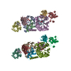
| ||||||||
|---|---|---|---|---|---|---|---|---|---|
| 1 | 
| ||||||||
| 2 | 
| ||||||||
| Unit cell |
| ||||||||
| Details | two heterooctameric molecules per asymmetric unit |
-Components
-Protein , 2 types, 8 molecules CGKODHLP
| #3: Protein | Mass: 11505.171 Da / Num. of mol.: 4 Source method: isolated from a genetically manipulated source Source: (gene. exp.) Paracoccus denitrificans (bacteria) / Production host: Rhodobacter sphaeroides (bacteria) / References: UniProt: P22364#4: Protein | Mass: 17094.650 Da / Num. of mol.: 4 Source method: isolated from a genetically manipulated source Source: (gene. exp.) Paracoccus denitrificans (bacteria) / Production host: Rhodobacter sphaeroides (bacteria) / References: UniProt: P29889, UniProt: P29899*PLUS |
|---|
-Methylamine dehydrogenase, ... / Antibody , 2 types, 8 molecules AEIMBFJN
| #1: Protein | Mass: 42673.492 Da / Num. of mol.: 4 / Mutation: alpha F55A of methylamine dehydrogenase Source method: isolated from a genetically manipulated source Source: (gene. exp.) Paracoccus denitrificans (bacteria) / Production host: Rhodobacter sphaeroides (bacteria) / References: UniProt: P29894, EC: 1.4.99.3#2: Antibody | Mass: 14210.696 Da / Num. of mol.: 4 Source method: isolated from a genetically manipulated source Source: (gene. exp.) Paracoccus denitrificans (bacteria) / Production host: Rhodobacter sphaeroides (bacteria) / References: UniProt: P22619, EC: 1.4.99.3 |
|---|
-Non-polymers , 5 types, 1705 molecules 


| #5: Chemical | ChemComp-PO4 /  Mass: 94.971 Da / Num. of mol.: 8 / Source method: obtained synthetically / Formula: PO4 Mass: 94.971 Da / Num. of mol.: 8 / Source method: obtained synthetically / Formula: PO4#6: Chemical | ChemComp-CU /  Mass: 63.546 Da / Num. of mol.: 4 / Source method: obtained synthetically / Formula: Cu Mass: 63.546 Da / Num. of mol.: 4 / Source method: obtained synthetically / Formula: Cu#7: Chemical | ChemComp-NA /  Mass: 22.990 Da / Num. of mol.: 4 / Source method: obtained synthetically / Formula: Na Mass: 22.990 Da / Num. of mol.: 4 / Source method: obtained synthetically / Formula: Na#8: Chemical | ChemComp-HEC /  Mass: 618.503 Da / Num. of mol.: 4 / Source method: obtained synthetically / Formula: C34H34FeN4O4 Mass: 618.503 Da / Num. of mol.: 4 / Source method: obtained synthetically / Formula: C34H34FeN4O4#9: Water | ChemComp-HOH / | Mass: 18.015 Da / Num. of mol.: 1685 / Source method: isolated from a natural source / Formula: H2O |
|---|
-Details
| Has protein modification | Y |
|---|
-Experimental details
-Experiment
| Experiment | Method: X-RAY DIFFRACTION / Number of used crystals: 1 |
|---|
- Sample preparation
| Crystal | Density Matthews: 2.73 Å3/Da / Density % sol: 54.94 % | ||||||||||||||||||||||||||||||||||||
|---|---|---|---|---|---|---|---|---|---|---|---|---|---|---|---|---|---|---|---|---|---|---|---|---|---|---|---|---|---|---|---|---|---|---|---|---|---|
| Crystal grow | Temperature: 295 K / Method: vapor diffusion, sitting drop / pH: 5 Details: phosphate, pH 5.0, VAPOR DIFFUSION, SITTING DROP, temperature 295K | ||||||||||||||||||||||||||||||||||||
| Crystal grow | *PLUS | ||||||||||||||||||||||||||||||||||||
| Components of the solutions | *PLUS
|
-Data collection
| Diffraction | Mean temperature: 100 K |
|---|---|
| Diffraction source | Source: ROTATING ANODE / Type: RIGAKU RU200 / Wavelength: 1.5418 Å |
| Detector | Type: RIGAKU RAXIS IIC / Detector: IMAGE PLATE / Date: Aug 6, 2001 |
| Radiation | Monochromator: GRAPHITE / Protocol: SINGLE WAVELENGTH / Monochromatic (M) / Laue (L): M / Scattering type: x-ray |
| Radiation wavelength | Wavelength: 1.5418 Å / Relative weight: 1 |
| Reflection | Resolution: 2.25→50 Å / Num. all: 173239 / Num. obs: 150718 / % possible obs: 87 % / Observed criterion σ(F): 0 / Observed criterion σ(I): 0 / Redundancy: 4.5 % / Biso Wilson estimate: 13.4 Å2 / Rmerge(I) obs: 0.074 / Net I/σ(I): 15.7 |
| Reflection shell | Resolution: 2.25→2.33 Å / Redundancy: 2.1 % / Rmerge(I) obs: 0.201 / Mean I/σ(I) obs: 4 / Num. unique all: 8212 / % possible all: 47.4 |
| Reflection shell | *PLUS Lowest resolution: 2.3 Å / % possible obs: 47.4 % |
- Processing
| Software |
| |||||||||||||||||||||||||||||||||||||||||||||||||||||||||||||||
|---|---|---|---|---|---|---|---|---|---|---|---|---|---|---|---|---|---|---|---|---|---|---|---|---|---|---|---|---|---|---|---|---|---|---|---|---|---|---|---|---|---|---|---|---|---|---|---|---|---|---|---|---|---|---|---|---|---|---|---|---|---|---|---|---|
| Refinement | Method to determine structure: MOLECULAR REPLACEMENT Starting model: PDB ENTRY 2MTA Resolution: 2.25→50 Å / Rfactor Rfree error: 0.002 / Isotropic thermal model: isotropic / Cross valid method: THROUGHOUT / σ(F): 0 / σ(I): 0 / Stereochemistry target values: Engh & Huber
| |||||||||||||||||||||||||||||||||||||||||||||||||||||||||||||||
| Solvent computation | Bsol: 35.5258 Å2 / ksol: 0.384412 e/Å3 | |||||||||||||||||||||||||||||||||||||||||||||||||||||||||||||||
| Displacement parameters | Biso mean: 22.8 Å2 | |||||||||||||||||||||||||||||||||||||||||||||||||||||||||||||||
| Refine analyze |
| |||||||||||||||||||||||||||||||||||||||||||||||||||||||||||||||
| Refinement step | Cycle: LAST / Resolution: 2.25→50 Å
| |||||||||||||||||||||||||||||||||||||||||||||||||||||||||||||||
| Refine LS restraints |
| |||||||||||||||||||||||||||||||||||||||||||||||||||||||||||||||
| LS refinement shell | Refine-ID: X-RAY DIFFRACTION / Total num. of bins used: 6
| |||||||||||||||||||||||||||||||||||||||||||||||||||||||||||||||
| Refinement | *PLUS % reflection Rfree: 10 % / Rfactor Rfree: 0.21 | |||||||||||||||||||||||||||||||||||||||||||||||||||||||||||||||
| Solvent computation | *PLUS | |||||||||||||||||||||||||||||||||||||||||||||||||||||||||||||||
| Displacement parameters | *PLUS | |||||||||||||||||||||||||||||||||||||||||||||||||||||||||||||||
| Refine LS restraints | *PLUS
| |||||||||||||||||||||||||||||||||||||||||||||||||||||||||||||||
| LS refinement shell | *PLUS Lowest resolution: 2.3 Å / Rfactor Rfree: 0.278 / Rfactor Rwork: 0.222 |
