 Movie
Movie Controller

[English] 日本語
 Yorodumi
- EMDB-6464: Reconstruction of the T20S proteasome at 2.8 Angstrom resolution ... -
+ Open data

- Basic information
| Entry | Database: EMDB / ID: EMD-6464 | |||||||||
|---|---|---|---|---|---|---|---|---|---|---|
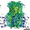
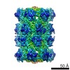
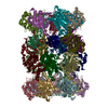
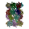
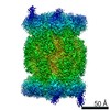
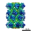
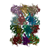
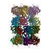
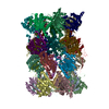
| Title | Reconstruction of the T20S proteasome at 2.8 Angstrom resolution using optimal exposure filtering | |||||||||
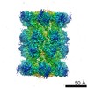
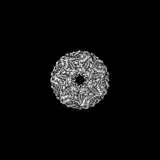
 Map data Map data | Unsharpened map of the T20S Proteasome obtained with exposure filtered particles and Frealign | |||||||||
 Sample Sample |
| |||||||||
 Keywords Keywords | exposure filter / dose / image processing | |||||||||
| Biological species |   Thermoplasma acidophilum (acidophilic) Thermoplasma acidophilum (acidophilic) | |||||||||
| Method | single particle reconstruction / cryo EM / Resolution: 2.8 Å | |||||||||
 Authors Authors | Grant T / Grigorieff N | |||||||||
 Citation Citation | Journal: Elife / Year: 2015 Title: 2.8 Å resolution reconstruction of the Thermoplasma acidophilum 20S proteasome using cryo-electron microscopy. Authors: Melody G Campbell / David Veesler / Anchi Cheng / Clinton S Potter / Bridget Carragher /  Abstract: Recent developments in detector hardware and image-processing software have revolutionized single particle cryo-electron microscopy (cryoEM) and led to a wave of near-atomic resolution (typically ...Recent developments in detector hardware and image-processing software have revolutionized single particle cryo-electron microscopy (cryoEM) and led to a wave of near-atomic resolution (typically ∼3.3 Å) reconstructions. Reaching resolutions higher than 3 Å is a prerequisite for structure-based drug design and for cryoEM to become widely interesting to pharmaceutical industries. We report here the structure of the 700 kDa Thermoplasma acidophilum 20S proteasome (T20S), determined at 2.8 Å resolution by single-particle cryoEM. The quality of the reconstruction enables identifying the rotameric conformation adopted by some amino-acid side chains (rotamers) and resolving ordered water molecules, in agreement with the expectations for crystal structures at similar resolutions. The results described in this manuscript demonstrate that single particle cryoEM is capable of competing with X-ray crystallography for determination of protein structures of suitable quality for rational drug design. | |||||||||
| History |
|
- Structure visualization
| Movie |
 Movie viewer Movie viewer |
|---|---|
| Structure viewer | EM map: SurfViewMolmilJmol/JSmol |
| Supplemental images |
- Downloads & links
-EMDB archive
| Map data | emd_6464.map.gz | 22.7 MB | EMDB map data format | |
|---|---|---|---|---|
| Header (meta data) | emd-6464-v30.xmlemd-6464.xml | 10.2 KB 10.2 KB | Display Display | EMDB header |
| Images |  400_6464.gif 400_6464.gif 80_6464.gif 80_6464.gif | 56.6 KB 4.4 KB | ||
| Archive directory |  http://ftp.pdbj.org/pub/emdb/structures/EMD-6464ftp://ftp.pdbj.org/pub/emdb/structures/EMD-6464 http://ftp.pdbj.org/pub/emdb/structures/EMD-6464ftp://ftp.pdbj.org/pub/emdb/structures/EMD-6464 | HTTPS FTP |
-Related structure data


-Links
| EMDB pages | EMDB (EBI/PDBe) / EMDataResource |
|---|
-Map
| File | Download / File: emd_6464.map.gz / Format: CCP4 / Size: 122.1 MB / Type: IMAGE STORED AS FLOATING POINT NUMBER (4 BYTES) | ||||||||||||||||||||||||||||||||||||||||||||||||||||||||||||||||||||
|---|---|---|---|---|---|---|---|---|---|---|---|---|---|---|---|---|---|---|---|---|---|---|---|---|---|---|---|---|---|---|---|---|---|---|---|---|---|---|---|---|---|---|---|---|---|---|---|---|---|---|---|---|---|---|---|---|---|---|---|---|---|---|---|---|---|---|---|---|---|
| Annotation | Unsharpened map of the T20S Proteasome obtained with exposure filtered particles and Frealign | ||||||||||||||||||||||||||||||||||||||||||||||||||||||||||||||||||||
| Projections & slices | Image control
Images are generated by Spider. | ||||||||||||||||||||||||||||||||||||||||||||||||||||||||||||||||||||
| Voxel size | X=Y=Z: 0.982 Å | ||||||||||||||||||||||||||||||||||||||||||||||||||||||||||||||||||||
| Density |
| ||||||||||||||||||||||||||||||||||||||||||||||||||||||||||||||||||||
| Symmetry | Space group: 1 | ||||||||||||||||||||||||||||||||||||||||||||||||||||||||||||||||||||
| Details | EMDB XML:
CCP4 map header:
| ||||||||||||||||||||||||||||||||||||||||||||||||||||||||||||||||||||
 Z (Sec.)
 Y (Row.)
 X (Col.)


-Supplemental data
- Sample components
-Entire : T20S Proteasome
| Entire | Name: T20S Proteasome |
|---|---|
| Components |
|
-Supramolecule #1000: T20S Proteasome
| Supramolecule | Name: T20S Proteasome / type: sample / ID: 1000 / Oligomeric state: D7 / Number unique components: 1 |
|---|---|
| Molecular weight | Theoretical: 700 KDa |
-Macromolecule #1: 20S proteasome
| Macromolecule | Name: 20S proteasome / type: protein_or_peptide / ID: 1 / Number of copies: 28 / Oligomeric state: 28-mer / Recombinant expression: Yes |
|---|---|
| Source (natural) | Organism: Thermoplasma acidophilum (acidophilic) |
| Molecular weight | Theoretical: 700 KDa |
| Recombinant expression | Organism:  |
-Experimental details
-Structure determination
| Method | cryo EM |
|---|---|
 Processing Processing | single particle reconstruction |
| Aggregation state | particle |
-Sample preparation
| Concentration | 0.21 mg/mL |
|---|---|
| Buffer | pH: 7.8 / Details: 20 mM Tris, 150 mM NaCl |
| Grid | Details: 1.2/1.3 C-Flat grid, plasma-cleaned |
| Vitrification | Cryogen name: ETHANE / Chamber temperature: 95 K / Instrument: GATAN CRYOPLUNGE 3 / Details: Vitrification carried out at room temperature. / Method: Blot for 2.5 seconds before plunging. |
- Electron microscopy
| Microscope | FEI TITAN KRIOS |
|---|---|
| Alignment procedure | Legacy - Astigmatism: Objective lens astigmatism was corrected at 22,500 times magnification. |
| Date | Sep 5, 2014 |
| Image recording | Category: CCD / Film or detector model: GATAN K2 (4k x 4k) / Number real images: 192 / Average electron dose: 53 e/Å2 Details: Each movie was acquired over 7.6 seconds and comprises 38 frames. |
| Electron beam | Acceleration voltage: 300 kV / Electron source:  FIELD EMISSION GUN FIELD EMISSION GUN |
| Electron optics | Calibrated magnification: 37313 / Illumination mode: FLOOD BEAM / Imaging mode: BRIGHT FIELD / Cs: 2.7 mm / Nominal defocus max: 2.4 µm / Nominal defocus min: 0.9 µm / Nominal magnification: 22500 |
| Sample stage | Specimen holder model: FEI TITAN KRIOS AUTOGRID HOLDER |
| Experimental equipment |  Model: Titan Krios / Image courtesy: FEI Company |
-Image processing
| Details | Particles were exposure-filtered then refined and reconstructed using Frealign. |
|---|---|
| CTF correction | Details: CTF parameters provided by Campbell et al. |
| Final reconstruction | Resolution.type: BY AUTHOR / Resolution: 2.8 Å / Resolution method: OTHER / Software - Name: Tigris, Unblur, Frealign Details: Map was created from exposure-filtered images. No information from resolutions greater than 5A was used in the refinement. Number images used: 49954 |
