 Movie
Movie Controller


+ Open data

- Basic information
| Entry | Database: PDB / ID: 1ivi | ||||||
|---|---|---|---|---|---|---|---|
| Title | Crystal Structure of pig dihydrolipoamide dehydrogenase | ||||||
 Components Components | dihydrolipoamide dehydrogenase | ||||||
 Keywords Keywords | OXIDOREDUCTASE / 2-OXOGLUTARATE DEHYDROGENASE COMPLEX / PYRUVATE DEHYDROGENASE COMPLEX | ||||||
| Biological species |  | ||||||
| Method |  X-RAY DIFFRACTION / SYNCHROTRON / Resolution: 8 Å X-RAY DIFFRACTION / SYNCHROTRON / Resolution: 8 Å | ||||||
 Authors Authors | Toyoda, T. / Kobayashi, R. / Sekiguchi, T. / Koike, K. / Koike, M. / Takenaka, A. | ||||||
 Citation Citation | Journal: Acta Crystallogr.,Sect.D / Year: 1998 Title: Crystallization and preliminary X-ray analysis of pig E3, lipoamide dehydrogenase. Authors: Toyoda, T. / Kobayashi, R. / Sekiguchi, T. / Koike, K. / Koike, M. / Takenaka, A. | ||||||
| History |
|
- Structure visualization
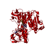
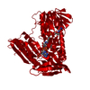
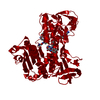
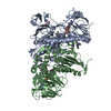
| Structure viewer | Molecule:  MolmilJmol/JSmol MolmilJmol/JSmol |
|---|
- Downloads & links
-Download
| PDBx/mmCIF format | 1ivi.cif.gz | 74.2 KB | Display | PDBx/mmCIF format |
|---|---|---|---|---|
| PDB format | pdb1ivi.ent.gz | 52.6 KB | Display | PDB format |
| PDBx/mmJSON format | 1ivi.json.gz | Tree view | PDBx/mmJSON format | |
| Others |  Other downloads Other downloads |
-Validation report
| Arichive directory | https://data.pdbj.org/pub/pdb/validation_reports/iv/1iviftp://data.pdbj.org/pub/pdb/validation_reports/iv/1ivi | HTTPS FTP |
|---|
-Related structure data
| Similar structure data |
|---|


-Links
 PDBj
PDBj
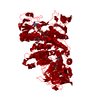
- Assembly
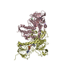
| Deposited unit | 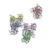
| ||||||||
|---|---|---|---|---|---|---|---|---|---|
| 1 | 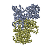
| ||||||||
| 2 | 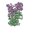
| ||||||||
| 3 | 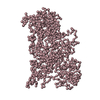
| ||||||||
| Unit cell |
|
-Components
| #1: Protein | Mass: 40698.164 Da / Num. of mol.: 5 / Source method: isolated from a natural source / Source: (natural) |
|---|
-Experimental details
-Experiment
| Experiment | Method: X-RAY DIFFRACTION / Number of used crystals: 1 |
|---|
- Sample preparation
| Crystal | Density Matthews: 6.43 Å3/Da / Density % sol: 80.88 % | ||||||||||||||||||||||||||||||||||||||||||
|---|---|---|---|---|---|---|---|---|---|---|---|---|---|---|---|---|---|---|---|---|---|---|---|---|---|---|---|---|---|---|---|---|---|---|---|---|---|---|---|---|---|---|---|
| Crystal grow | Temperature: 298 K / Method: vapor diffusion, hanging drop / pH: 7 Details: Ammonium sulphate, pH 7.0, VAPOR DIFFUSION, HANGING DROP, temperature 298K | ||||||||||||||||||||||||||||||||||||||||||
| Crystal grow | *PLUS | ||||||||||||||||||||||||||||||||||||||||||
| Components of the solutions | *PLUS
|
-Data collection
| Diffraction | Mean temperature: 293 K |
|---|---|
| Diffraction source | Source: SYNCHROTRON / Site: Photon Factory  / Beamline: BL-6A / Wavelength: 1.04 Å / Beamline: BL-6A / Wavelength: 1.04 Å |
| Detector | Type: FUJI / Detector: IMAGE PLATE / Details: mirrors |
| Radiation | Monochromator: graphite / Protocol: SINGLE WAVELENGTH / Monochromatic (M) / Laue (L): M / Scattering type: x-ray |
| Radiation wavelength | Wavelength: 1.04 Å / Relative weight: 1 |
| Reflection | Resolution: 8→50 Å / Num. all: 13615 / Num. obs: 5838 / Rmerge(I) obs: 0.066 |
| Reflection | *PLUS Highest resolution: 8 Å / % possible obs: 98 % / Num. measured all: 13615 |
- Processing
| Software |
| |||||||||||||||
|---|---|---|---|---|---|---|---|---|---|---|---|---|---|---|---|---|
| Refinement | Resolution: 8→50 Å / Rfactor Rwork: 0.378 Details: The coordinates for only the alpha carbons are present in the structure. | |||||||||||||||
| Refinement step | Cycle: LAST / Resolution: 8→50 Å
| |||||||||||||||
| Refinement | *PLUS Lowest resolution: 50 Å / % reflection Rfree: 10 % / Rfactor Rfree: 0.379 | |||||||||||||||
| Solvent computation | *PLUS | |||||||||||||||
| Displacement parameters | *PLUS |
