ムービー
ムービー コントローラー


+ データを開く

- 基本情報
基本情報
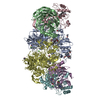
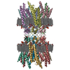
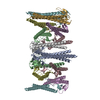
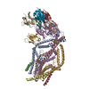
| 登録情報 | データベース: PDB / ID: 3j6c | ||||||
|---|---|---|---|---|---|---|---|
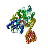
| タイトル | Cryo-EM structure of MAVS CARD filament | ||||||
 要素 要素 | Mitochondrial antiviral-signaling protein | ||||||
 キーワード キーワード | SIGNALING PROTEIN / Innate immunity / helical filament | ||||||
| 機能・相同性 |  機能・相同性情報 機能・相同性情報positive regulation of IP-10 production / regulation of peroxisome organization / positive regulation of chemokine (C-C motif) ligand 5 production / positive regulation of myeloid dendritic cell cytokine production / CARD domain binding / NF-kB activation through FADD/RIP-1 pathway mediated by caspase-8 and -10 / positive regulation of response to cytokine stimulus / protein localization to mitochondrion / positive regulation of type I interferon-mediated signaling pathway / peroxisomal membrane ...positive regulation of IP-10 production / regulation of peroxisome organization / positive regulation of chemokine (C-C motif) ligand 5 production / positive regulation of myeloid dendritic cell cytokine production / CARD domain binding / NF-kB activation through FADD/RIP-1 pathway mediated by caspase-8 and -10 / positive regulation of response to cytokine stimulus / protein localization to mitochondrion / positive regulation of type I interferon-mediated signaling pathway / peroxisomal membrane / TRAF6 mediated IRF7 activation / negative regulation of viral genome replication / cytoplasmic pattern recognition receptor signaling pathway / type I interferon-mediated signaling pathway / cellular response to exogenous dsRNA / positive regulation of NLRP3 inflammasome complex assembly / TRAF6 mediated NF-kB activation / positive regulation of interferon-alpha production / positive regulation of type I interferon production / ubiquitin ligase complex / positive regulation of defense response to virus by host / signaling adaptor activity / antiviral innate immune response / activation of innate immune response / protein serine/threonine kinase binding / positive regulation of interferon-beta production / Negative regulators of DDX58/IFIH1 signaling / positive regulation of interleukin-8 production / DDX58/IFIH1-mediated induction of interferon-alpha/beta / Evasion by RSV of host interferon responses / molecular condensate scaffold activity / mitochondrial membrane / PKR-mediated signaling / positive regulation of interleukin-6 production / positive regulation of protein import into nucleus / SARS-CoV-1 activates/modulates innate immune responses / Ovarian tumor domain proteases / positive regulation of tumor necrosis factor production / TRAF3-dependent IRF activation pathway / protein-macromolecule adaptor activity / defense response to virus / molecular adaptor activity / DNA-binding transcription factor binding / mitochondrial outer membrane / positive regulation of canonical NF-kappaB signal transduction / intracellular signal transduction / defense response to bacterium / innate immune response / protein kinase binding / SARS-CoV-2 activates/modulates innate and adaptive immune responses / signal transduction / positive regulation of transcription by RNA polymerase II / mitochondrion / identical protein binding 類似検索 - 分子機能 | ||||||
| 生物種 |  Homo sapiens (ヒト) Homo sapiens (ヒト) | ||||||
| 手法 | 電子顕微鏡法 / らせん対称体再構成法 / クライオ電子顕微鏡法 / 解像度: 9.6 Å | ||||||
 データ登録者 データ登録者 | Xu, H. / He, X. / Zheng, H. / Huang, L.J. / Hou, F. / Yu, Z. / de la Cruz, M.J. / Borkowski, B. / Zhang, X. / Chen, Z.J. / Jiang, Q.-X. | ||||||
 引用 引用 | ジャーナル: Elife / 年: 2014 タイトル: Structural basis for the prion-like MAVS filaments in antiviral innate immunity. 著者: Hui Xu / Xiaojing He / Hui Zheng / Lily J Huang / Fajian Hou / Zhiheng Yu / Michael Jason de la Cruz / Brian Borkowski / Xuewu Zhang / Zhijian J Chen / Qiu-Xing Jiang /  要旨: Mitochondrial antiviral signaling (MAVS) protein is required for innate immune responses against RNA viruses. In virus-infected cells MAVS forms prion-like aggregates to activate antiviral signaling ...Mitochondrial antiviral signaling (MAVS) protein is required for innate immune responses against RNA viruses. In virus-infected cells MAVS forms prion-like aggregates to activate antiviral signaling cascades, but the underlying structural mechanism is unknown. Here we report cryo-electron microscopic structures of the helical filaments formed by both the N-terminal caspase activation and recruitment domain (CARD) of MAVS and a truncated MAVS lacking part of the proline-rich region and the C-terminal transmembrane domain. Both structures are left-handed three-stranded helical filaments, revealing specific interfaces between individual CARD subunits that are dictated by electrostatic interactions between neighboring strands and hydrophobic interactions within each strand. Point mutations at multiple locations of these two interfaces impaired filament formation and antiviral signaling. Super-resolution imaging of virus-infected cells revealed rod-shaped MAVS clusters on mitochondria. These results elucidate the structural mechanism of MAVS polymerization, and explain how an α-helical domain uses distinct chemical interactions to form self-perpetuating filaments. DOI: http://dx.doi.org/10.7554/eLife.01489.001. #1: ジャーナル: BMC Struct Biol / 年: 2008タイトル: Crystal structure of human IPS-1/MAVS/VISA/Cardif caspase activation recruitment domain. 著者: Jane A Potter / Richard E Randall / Garry L Taylor /  要旨: BACKGROUND: IPS-1/MAVS/VISA/Cardif is an adaptor protein that plays a crucial role in the induction of interferons in response to viral infection. In the initial stage of the intracellular antiviral ...BACKGROUND: IPS-1/MAVS/VISA/Cardif is an adaptor protein that plays a crucial role in the induction of interferons in response to viral infection. In the initial stage of the intracellular antiviral response two RNA helicases, retinoic acid inducible gene-I (RIG-I) and melanoma differentiation-association gene 5 (MDA5), are independently able to bind viral RNA in the cytoplasm. The 62 kDa protein IPS-1/MAVS/VISA/Cardif contains an N-terminal caspase activation and recruitment (CARD) domain that associates with the CARD regions of RIG-I and MDA5, ultimately leading to the induction of type I interferons. As a first step towards understanding the molecular basis of this important adaptor protein we have undertaken structural studies of the IPS-1 MAVS/VISA/Cardif CARD region. RESULTS: The crystal structure of human IPS-1/MAVS/VISA/Cardif CARD has been determined to 2.1A resolution. The protein was expressed and crystallized as a maltose-binding protein (MBP) fusion ...RESULTS: The crystal structure of human IPS-1/MAVS/VISA/Cardif CARD has been determined to 2.1A resolution. The protein was expressed and crystallized as a maltose-binding protein (MBP) fusion protein. The MBP and IPS-1 components each form a distinct domain within the structure. IPS-1/MAVS/VISA/Cardif CARD adopts a characteristic six-helix bundle with a Greek-key topology and, in common with a number of other known CARD structures, contains two major polar surfaces on opposite sides of the molecule. One face has a surface-exposed, disordered tryptophan residue that may explain the poor solubility of untagged expression constructs. CONCLUSION: The IPS-1/MAVS/VISA/Cardif CARD domain adopts the classic CARD fold with an asymmetric surface charge distribution that is typical of CARD domains involved in homotypic protein-protein ...CONCLUSION: The IPS-1/MAVS/VISA/Cardif CARD domain adopts the classic CARD fold with an asymmetric surface charge distribution that is typical of CARD domains involved in homotypic protein-protein interactions. The location of the two polar areas on IPS-1/MAVS/VISA/Cardif CARD suggest possible types of associations that this domain makes with the two CARD domains of MDA5 or RIG-I. The N-terminal CARD domains of RIG-I and MDA5 share greatest sequence similarity with IPS-1/MAVS/VISA/Cardif CARD and this has allowed modelling of their structures. These models show a very different charge profile for the equivalent surfaces compared to IPS-1/MAVS/VISA/Cardif CARD. | ||||||
| 履歴 |
|
- 構造の表示
構造の表示
| ムービー |
ムービービューア |
|---|---|
| 構造ビューア | 分子: MolmilJmol/JSmol |
- ダウンロードとリンク
-ダウンロード
| PDBx/mmCIF形式 | 3j6c.cif.gz | 31.2 KB | 表示 | PDBx/mmCIF形式 |
|---|---|---|---|---|
| PDB形式 | pdb3j6c.ent.gz | 20.3 KB | 表示 | PDB形式 |
| PDBx/mmJSON形式 | 3j6c.json.gz | ツリー表示 | PDBx/mmJSON形式 | |
| その他 |  その他のダウンロード その他のダウンロード |
-検証レポート
| 文書・要旨 | 3j6c_validation.pdf.gz | 695.7 KB | 表示 | wwPDB検証レポート |
|---|---|---|---|---|
| 文書・詳細版 | 3j6c_full_validation.pdf.gz | 695.4 KB | 表示 | |
| XML形式データ | 3j6c_validation.xml.gz | 9.1 KB | 表示 | |
| CIF形式データ | 3j6c_validation.cif.gz | 12.1 KB | 表示 | |
| アーカイブディレクトリ | https://data.pdbj.org/pub/pdb/validation_reports/j6/3j6cftp://data.pdbj.org/pub/pdb/validation_reports/j6/3j6c | HTTPS FTP |
-関連構造データ
| 関連構造データ |  5890MC 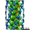 5891C 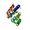 4o9fC 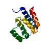 4o9lC M: このデータのモデリングに利用したマップデータ C: 同じ文献を引用 ( |
|---|---|
| 類似構造データ | |
| 電子顕微鏡画像生データ | EMPIAR-10014 (タイトル: MAVS CARD and DeltaProTM filaments / Data size: 35.3 / Data #1: CARD set1 micrographs [micrographs - single frame] / Data #2: CARD set2 micrographs [micrographs - single frame] / Data #3: CARD set3 micrographs [micrographs - single frame] / Data #4: CARD set4 micrographs [micrographs - single frame] Data #5: CARD set1 segments [picked particles - multiframe - unprocessed] Data #6: CARD set2 segments [picked particles - multiframe - unprocessed] Data #7: CARD set3 segments [picked particles - multiframe - unprocessed] Data #8: CARD set4 segments [picked particles - multiframe - unprocessed] Data #9: DeltProTM micrographs [micrographs - single frame] Data #10: DeltaProTM segments [picked particles - multiframe - unprocessed]) |


-リンク
 PDBj
PDBj


- 集合体
集合体
| 登録構造単位 | 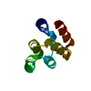
|
|---|---|
| 1 | x 24
|
| 2 |
|
| 3 | 
|
| 対称性 | らせん対称: (回転対称性: 3 / Dyad axis: no / N subunits divisor: 1 / Num. of operations: 24 / Rise per n subunits: 16.8 Å / Rotation per n subunits: -53.6 °) |
-要素
| #1: タンパク質 | 分子量: 10826.325 Da / 分子数: 1 断片: caspase activation recruitment domain (UNP residues 3-93) 由来タイプ: 組換発現 / 由来: (組換発現) Homo sapiens (ヒト) / 遺伝子: MAVS, IPS1, KIAA1271, VISA / プラスミド: pcDNA3 / 細胞株 (発現宿主): HEK293T / 発現宿主: Homo sapiens (ヒト) / 参照: UniProt: Q7Z434 |
|---|
-実験情報
-実験
| 実験 | 手法: 電子顕微鏡法 |
|---|---|
| EM実験 | 試料の集合状態: FILAMENT / 3次元再構成法: らせん対称体再構成法 |
- 試料調製
試料調製
| 構成要素 | 名称: MAVS CARD filament / タイプ: COMPLEX / 詳細: polymer |
|---|---|
| 分子量 | 値: 0.546 MDa / 実験値: NO |
| 緩衝液 | 名称: 20 mM Tris-HCl, 50 mM NaCl, 1 mM DTT / pH: 7.5 / 詳細: 20 mM Tris-HCl, 50 mM NaCl, 1 mM DTT |
| 試料 | 包埋: NO / シャドウイング: NO / 染色: NO / 凍結: YES |
| 試料支持 | 詳細: Quantifoil grids coated with thin carbon on top |
| 急速凍結 | 装置: FEI VITROBOT MARK III / 凍結剤: ETHANE / 湿度: 100 % / 詳細: Plunged into liquid ethane (FEI VITROBOT MARK III) |
- 電子顕微鏡撮影
| 顕微鏡 | モデル: JEOL 2200FS / 日付: 2011年10月20日 |
|---|---|
| 電子銃 | 電子線源:  FIELD EMISSION GUN / 加速電圧: 200 kV / 照射モード: FLOOD BEAM FIELD EMISSION GUN / 加速電圧: 200 kV / 照射モード: FLOOD BEAM |
| 電子レンズ | モード: BRIGHT FIELD / 倍率(公称値): 60000 X / 倍率(補正後): 61950 X / 最大 デフォーカス(公称値): 2000 nm / 最小 デフォーカス(公称値): 800 nm / Cs: 2 mm 非点収差: Objective lens astigmatism was corrected at 100,000x magnification カメラ長: 0 mm |
| 試料ホルダ | 試料ホルダーモデル: GATAN LIQUID NITROGEN / 傾斜角・最大: 0 ° / 傾斜角・最小: 0 ° |
| 撮影 | 電子線照射量: 20 e/Å2 / フィルム・検出器のモデル: KODAK SO-163 FILM |
| 電子光学装置 | エネルギーフィルター名称: FEI / エネルギーフィルター 上限: 35 eV / エネルギーフィルター 下限: 0 eV |
| 放射 | プロトコル: SINGLE WAVELENGTH / 単色(M)・ラウエ(L): M / 散乱光タイプ: x-ray |
| 放射波長 | 相対比: 1 |
- 解析
解析
| EMソフトウェア |
| ||||||||||||||||||||||||||||
|---|---|---|---|---|---|---|---|---|---|---|---|---|---|---|---|---|---|---|---|---|---|---|---|---|---|---|---|---|---|
| CTF補正 | 詳細: Each filament segment | ||||||||||||||||||||||||||||
| らせん対称 | 回転角度/サブユニット: 53.6 ° / 軸方向距離/サブユニット: 16.8 Å / らせん対称軸の対称性: C3 | ||||||||||||||||||||||||||||
| 3次元再構成 | 手法: IHRSR / 解像度: 9.6 Å / 解像度の算出法: FSC 0.5 CUT-OFF / ピクセルサイズ(公称値): 2.333 Å / ピクセルサイズ(実測値): 2.333 Å 詳細: Final data were calculated from three separate datasets from three sessions of data collection. The handedness of the map was determined by cryo-electron tomography (Helical Details: IHRSR). 対称性のタイプ: HELICAL | ||||||||||||||||||||||||||||
| 原子モデル構築 | プロトコル: RIGID BODY FIT / 空間: REAL 詳細: REFINEMENT PROTOCOL--rigid body DETAILS--The docking of one X-ray model into a segmented map corresponding to one subunit was first done manually in Chimera, and then optimized using SITUS. | ||||||||||||||||||||||||||||
| 原子モデル構築 | PDB-ID: 2VGQ PDB chain-ID: A / Accession code: 2VGQ / Source name: PDB / タイプ: experimental model | ||||||||||||||||||||||||||||
| 精密化ステップ | サイクル: LAST
|