[English] 日本語
Yorodumi
- PDB-4o9l: crystal structure of horse MAVS card domain mutant E26R -

+
Open data


ID or keywords:

Loading...

-
Basic information

Entry
Database: PDB / ID: 4o9l
Titlecrystal structure of horse MAVS card domain mutant E26R
Componentsmitochondrial antiviral signaling protein (MAVS)
KeywordsANTIVIRAL PROTEIN
Function / homology
Function and homology information


defense response to virus / membrane => GO:0016020 / mitochondrion
Similarity search - Function
Mitochondrial antiviral-signalling protein, metazoan / Death Domain, Fas / Caspase recruitment domain / Caspase recruitment domain / Death Domain, Fas / Orthogonal Bundle / Mainly Alpha
Similarity search - Domain/homology
Mitochondrial antiviral signaling protein
Similarity search - Component
Biological speciesEquus caballus (horse)
MethodX-RAY DIFFRACTION / SYNCHROTRON / MOLECULAR REPLACEMENT / Resolution: 1.944 Å
AuthorsZhang, X. / He, X.
Citation
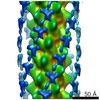
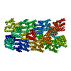
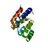
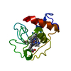
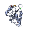
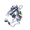
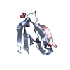
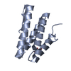
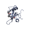
Journal: Elife / Year: 2014
Title: Structural basis for the prion-like MAVS filaments in antiviral innate immunity.
Authors: Hui Xu / Xiaojing He / Hui Zheng / Lily J Huang / Fajian Hou / Zhiheng Yu / Michael Jason de la Cruz / Brian Borkowski / Xuewu Zhang / Zhijian J Chen / Qiu-Xing Jiang /
Abstract: Mitochondrial antiviral signaling (MAVS) protein is required for innate immune responses against RNA viruses. In virus-infected cells MAVS forms prion-like aggregates to activate antiviral signaling ...Mitochondrial antiviral signaling (MAVS) protein is required for innate immune responses against RNA viruses. In virus-infected cells MAVS forms prion-like aggregates to activate antiviral signaling cascades, but the underlying structural mechanism is unknown. Here we report cryo-electron microscopic structures of the helical filaments formed by both the N-terminal caspase activation and recruitment domain (CARD) of MAVS and a truncated MAVS lacking part of the proline-rich region and the C-terminal transmembrane domain. Both structures are left-handed three-stranded helical filaments, revealing specific interfaces between individual CARD subunits that are dictated by electrostatic interactions between neighboring strands and hydrophobic interactions within each strand. Point mutations at multiple locations of these two interfaces impaired filament formation and antiviral signaling. Super-resolution imaging of virus-infected cells revealed rod-shaped MAVS clusters on mitochondria. These results elucidate the structural mechanism of MAVS polymerization, and explain how an α-helical domain uses distinct chemical interactions to form self-perpetuating filaments. DOI: http://dx.doi.org/10.7554/eLife.01489.001.
History
DepositionJan 2, 2014Deposition site: RCSB / Processing site: RCSB
Revision 1.0Mar 12, 2014Provider: repository / Type: Initial release
Revision 1.1Nov 22, 2017Group: Refinement description / Category: software / Item: _software.name
Revision 1.2Sep 20, 2023Group: Data collection / Database references / Refinement description
Category: chem_comp_atom / chem_comp_bond ...chem_comp_atom / chem_comp_bond / database_2 / pdbx_initial_refinement_model / struct_ref_seq_dif
Item: _database_2.pdbx_DOI / _database_2.pdbx_database_accession / _struct_ref_seq_dif.details

-
Structure visualization

Structure viewerMolecule:
MolmilJmol/JSmol

Downloads & links

-
Assembly

Deposited unit
A: mitochondrial antiviral signaling protein (MAVS)


Theoretical massNumber of molelcules
Total (without water)11,3291
Polymers11,3291
Non-polymers00
Water2,810156
1


  • Idetical with deposited unit
  • defined by author&software
TypeNameSymmetry operationNumber
identity operation1_555x,y,z1
MethodPISA
Unit cell
Length a, b, c (Å)50.614, 51.942, 35.754
Angle α, β, γ (deg.)90.00, 90.00, 90.00
Int Tables number18
Space group name H-MP21212
Components on special symmetry positions
IDModelComponents
11A-177-

HOH

21A-212-

HOH

-
Components

#1: Protein mitochondrial antiviral signaling protein (MAVS)


Mass: 11328.824 Da / Num. of mol.: 1 / Fragment: card domain, UNP residues 1-94 / Mutation: E26R
Source method: isolated from a genetically manipulated source
Source: (gene. exp.) Equus caballus (horse) / Gene: MAVS / Plasmid: modified pET28a with a preScission protease site / Production host: Escherichia coli (E. coli) / Strain (production host): BL21(DE3) / References: UniProt: F6QPU3
#2: Water ChemComp-HOH / water / Water


Mass: 18.015 Da / Num. of mol.: 156 / Source method: isolated from a natural source / Formula: H2O

-
Experimental details

-
Experiment

ExperimentMethod: X-RAY DIFFRACTION / Number of used crystals: 1

-
Sample preparation

CrystalDensity Matthews: 2.07 Å3/Da / Density % sol: 40.7 %
Crystal growTemperature: 293 K / Method: vapor diffusion, hanging drop / pH: 6.5
Details: 30% PEGMME 3350, 0.2M ammonium sulfate, 0.1M Mes, pH 6.5, VAPOR DIFFUSION, HANGING DROP, temperature 293K

-
Data collection

DiffractionMean temperature: 200 K
Diffraction sourceSource: SYNCHROTRON / Site: APS / Beamline: 19-ID / Wavelength: 0.97926 Å
DetectorType: ADSC QUANTUM 315r / Detector: CCD / Date: Jun 13, 2012
RadiationMonochromator: Si 111 CHANNEL / Protocol: SINGLE WAVELENGTH / Monochromatic (M) / Laue (L): M / Scattering type: x-ray
Radiation wavelengthWavelength: 0.97926 Å / Relative weight: 1
ReflectionResolution: 1.8→50 Å / Num. all: 9177 / Num. obs: 8395 / % possible obs: 91.5 % / Observed criterion σ(F): 0 / Observed criterion σ(I): 0
Reflection shellResolution: 1.8→1.83 Å / % possible all: 55

-
Processing

Software
NameVersionClassification
HKL-3000data collection
CCP4model building
PHENIX(phenix.refine: 1.8.2_1309)refinement
HKL-3000data reduction
HKL-3000data scaling
CCP4phasing
RefinementMethod to determine structure: MOLECULAR REPLACEMENT
Starting model: PDB ENTRY 2VGQ
Resolution: 1.944→29.203 Å / SU ML: 0.19 / Isotropic thermal model: ISOTROPIC / Cross valid method: THROUGHOUT / σ(F): 1.37 / Phase error: 20.2 / Stereochemistry target values: ML
RfactorNum. reflection% reflectionSelection details
Rfree0.2092 332 4.61 %RANDOM
Rwork0.1625 ---
obs0.1646 7206 97.87 %-
all-7538 --
Solvent computationShrinkage radii: 0.9 Å / VDW probe radii: 1.11 Å / Solvent model: FLAT BULK SOLVENT MODEL
Refinement stepCycle: LAST / Resolution: 1.944→29.203 Å
ProteinNucleic acidLigandSolventTotal
Num. atoms788 0 0 156 944
Refine LS restraints
Refine-IDTypeDev idealNumber
X-RAY DIFFRACTIONf_bond_d0.007805
X-RAY DIFFRACTIONf_angle_d0.9431089
X-RAY DIFFRACTIONf_dihedral_angle_d13.521295
X-RAY DIFFRACTIONf_chiral_restr0.068121
X-RAY DIFFRACTIONf_plane_restr0.003138
LS refinement shell

Refine-ID: X-RAY DIFFRACTION

Resolution (Å)Rfactor RfreeNum. reflection RfreeRfactor RworkNum. reflection RworkNum. reflection obs% reflection obs (%)
1.9436-2.44860.2261540.16643324332496
2.4486-29.2060.20031780.16053550355099
Refinement TLS params.Method: refined / Origin x: 5.8373 Å / Origin y: 9.6041 Å / Origin z: 8.9739 Å
111213212223313233
T0.0475 Å20.0005 Å20.0132 Å2-0.0317 Å20 Å2--0.03 Å2
L1.763 °20.5817 °20.5817 °2-0.3039 °20.2069 °2--0.5825 °2
S-0.0132 Å °0.0306 Å °0.0695 Å °-0.0405 Å °-0.008 Å °-0.0127 Å °-0.0357 Å °0.0386 Å °-0.0042 Å °
Refinement TLS groupSelection details: chain A

+
About Yorodumi

-
News

-
Feb 9, 2022. New format data for meta-information of EMDB entries

New format data for meta-information of EMDB entries

  • Version 3 of the EMDB header file is now the official format.
  • The previous official version 1.9 will be removed from the archive.

Related info.:EMDB header

External links:wwPDB to switch to version 3 of the EMDB data model

-
Aug 12, 2020. Covid-19 info

Covid-19 info

URL: https://pdbj.org/emnavi/covid19.php

New page: Covid-19 featured information page in EM Navigator.

Related info.:Covid-19 info / Mar 5, 2020. Novel coronavirus structure data

+
Mar 5, 2020. Novel coronavirus structure data

Novel coronavirus structure data

Related info.:Yorodumi Speices / Aug 12, 2020. Covid-19 info

External links:COVID-19 featured content - PDBj / Molecule of the Month (242):Coronavirus Proteases

+
Jan 31, 2019. EMDB accession codes are about to change! (news from PDBe EMDB page)

EMDB accession codes are about to change! (news from PDBe EMDB page)

  • The allocation of 4 digits for EMDB accession codes will soon come to an end. Whilst these codes will remain in use, new EMDB accession codes will include an additional digit and will expand incrementally as the available range of codes is exhausted. The current 4-digit format prefixed with “EMD-” (i.e. EMD-XXXX) will advance to a 5-digit format (i.e. EMD-XXXXX), and so on. It is currently estimated that the 4-digit codes will be depleted around Spring 2019, at which point the 5-digit format will come into force.
  • The EM Navigator/Yorodumi systems omit the EMD- prefix.

Related info.:Q: What is EMD? / ID/Accession-code notation in Yorodumi/EM Navigator

External links:EMDB Accession Codes are Changing Soon! / Contact to PDBj

+
Jul 12, 2017. Major update of PDB

Major update of PDB

  • wwPDB released updated PDB data conforming to the new PDBx/mmCIF dictionary.
  • This is a major update changing the version number from 4 to 5, and with Remediation, in which all the entries are updated.
  • In this update, many items about electron microscopy experimental information are reorganized (e.g. em_software).
  • Now, EM Navigator and Yorodumi are based on the updated data.

External links:wwPDB Remediation / Enriched Model Files Conforming to OneDep Data Standards Now Available in the PDB FTP Archive

-
Yorodumi

Thousand views of thousand structures

  • Yorodumi is a browser for structure data from EMDB, PDB, SASBDB, etc.
  • This page is also the successor to EM Navigator detail page, and also detail information page/front-end page for Omokage search.
  • The word "yorodu" (or yorozu) is an old Japanese word meaning "ten thousand". "mi" (miru) is to see.

Related info.:EMDB / PDB / SASBDB / Comparison of 3 databanks / Yorodumi Search / Aug 31, 2016. New EM Navigator & Yorodumi / Yorodumi Papers / Jmol/JSmol / Function and homology information / Changes in new EM Navigator and Yorodumi

Read more